Ernest Hinton, 1853 – 1909
by Brian Stevenson
last updated April, 2020
Ernest Hinton was a full-time,
professional microscope slide preparer, working from 1864 through the turn of
the century. His high quality mounts are readily identified by his small, neat
handwriting (Figure 1). Hinton was reported to have made a large proportion of
Edmund Wheeler’s output of slides, which is probably true. After Wheeler’s
retirement, Hinton sold his wares from his own shop and through the C. Baker
microscopy company (Figure 1). In addition to slides, he manufactured various apparatus for the microscope, such as lenses, lamps, condensers, and, apparently, entire microscopes. Hinton’s personal life was full of tragedy: his father died when Ernest was young, forcing the family into poverty and Ernest to early labor, his wife died while she was still young, and Ernest himself died a painful death from cancer.
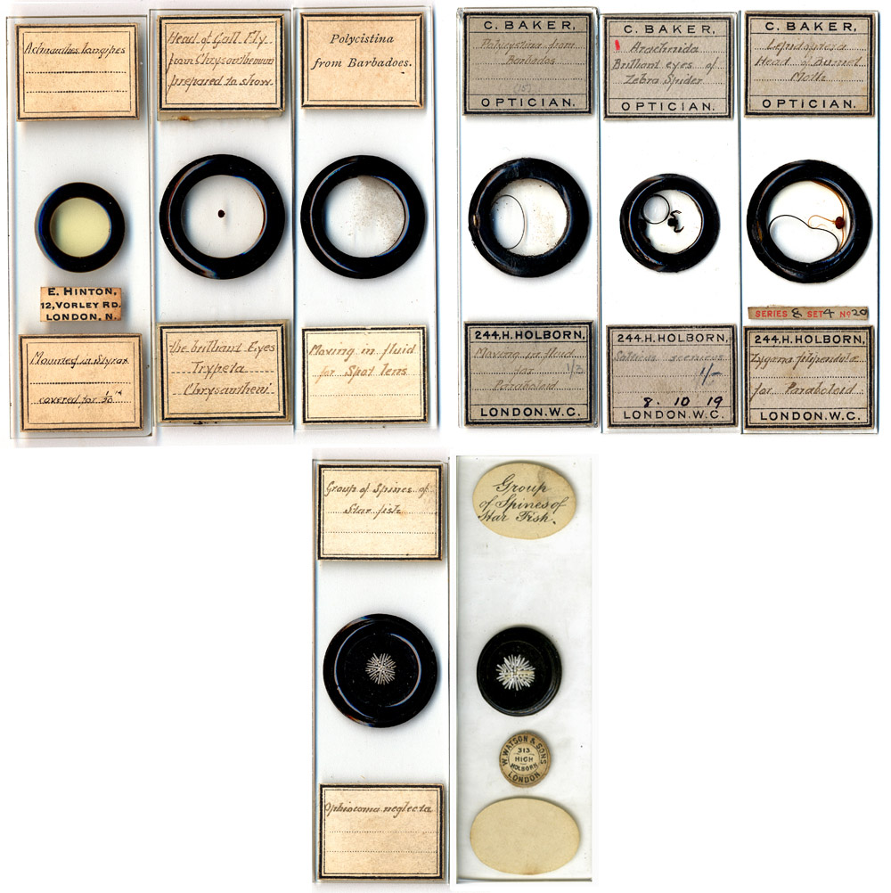
Figure 1. Examples of microscope slides made by Ernest Hinton, with his own labels (top row, right) and those of Baker (top row, left). Two identical mounts of Barbados polycistina floating in fluid are shown, one with each type of label. Hinton exhibited a slide of the head of a gall fly (Tryptera chrysanthemi) to the Royal Microscopical Society in 1897 (top row, second from left). The bottom row shows an arrangement of starfish spines, with Hinton labels, and a nearly identical slide with specimen description by Wheeler and a Watson and Sons retail label. Wheeler sold his stock to Watson in 1884, who then added their circular trade labels. The similarities between the Hinton and Wheeler/Watson slides suggest that Ernest Hinton made both of them, after and before 1884, respectively. A magnified view of the Hinton starfish spines is shown in Figure 6, at the end of this essay.
Ernest Hinton was born during
the summer of 1853, the second (and last) child of George and Sarah Hinton.
George was an artist, as was also his father. The workmanship of Ernest’s
microscopical mounts indicates that the family tradition was passed on to him.
George described himself as a “gilder and
bronzer” for the 1851 census, as a worker in a “fancy repository” for the 1861 census, and as an “artist” on his 1845 marriage record. The
Hinton’s were moderately well-off, recorded as employing a live-in house
servant on both the 1851 and 1861 censuses. During 1851, the family lived in a
private home at 10 Camford Terrace, St Pancras, Middlesex, and in 1861 at 14
High Street, Hampstead, Middlesex.
Life changed dramatically when
Ernest’s father died in February, 1864. The 1871 and 1881 censuses report
Ernest and his mother, Sarah, sharing houses with other families. There were no
servants. Sarah went to work, in 1871 being reported to be a “machinist”. 11-year old Ernest dropped
out of school, and went to work. Ernest’s later advertisements stated that
he worked for Edmund Wheeler for 20 years (Figure 2). Wheeler sold his business in 1884,
just before his death. That indicates that Hinton began working for Wheeler
right after his father’s death. At that time, the Hintons lived at 42 Grafton
Rd, an 8 minute walk from Wheeler’s home and business at 48 Tollington Rd.
Hinton’s artistic background may have be particularly attractive to Wheeler,
inducing him to hire the young orphan. It is unclear whether or not Wheeler
would have known the Hintons socially. Wheeler was a Quaker (Society of Friends), as was also his other known employee, nephew Frederick
Enock. George and Sarah Hinton were married at St. Paul’s, Canonbury, following
Anglican rites. At some point between 1871 and 1881, Ernest and his mother moved to 12 Vorley
Rd., a 4-5 minute walk to work at Wheeler’s shop. In 1881, the Hintons shared
this house with an unrelated couple, John and Isabella Laurence, from Chester.
Ernest remained at that house for most of the remainder of his life, although
without co-tenants. Edmund Wheeler’s retirement in 1884, and the resulting
change in Ernest Hinton’s position from assistant to business owner undoubtedly
improved his financial status. The Hintons never again had a house servant, though.
In 1882, Ernest married Clara
Moir. Census records and Clara’s death record indicate that the couple lived
with Ernest’s mother, Sarah, at 12 Vorley Rd. Two years after their marriage,
Clara was diagnosed with breast cancer. She died 5 years later, in November,
1889. Ernest and Clara did not have any children.
I did not find any
advertisements or other mentions of Hinton as a microscopist prior to late
1884, suggesting that he worked with Wheeler until the employer closed his
business. Almost immediately after Wheeler’s retirement, large advertisements
from Hinton appeared regularly in Science-Gossip and other popular magazines (Figure 2). Hinton’s 20 years of experience working
with Wheeler was stressed, even long after the old man was dead, attesting to
Wheeler’s enduring reputation for quality workmanship.
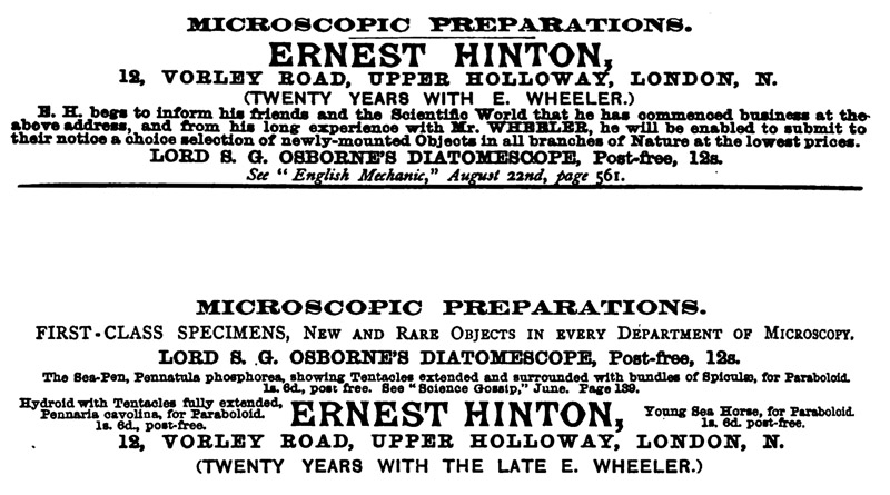
Figure 2.
Advertisements from Ernest Hinton that appeared in Hardwicke’s Science-Gossip
during 1885 (upper) and 1892 (lower).
Hinton quickly established a working relationship with Lord
Sidney Godolphin Osborne, a noted microscopist. Osborne had invented a
condenser that could be attached to the stage of any microscope, to enhance
illumination when viewing diatoms. Osborne called his devise the “Diatomescope”
(Figure 3). This device was distinct from a portable, hand-held diatom-viewer
to which he gave the same name (see Giordano’s Singular Beauty, page 59, for a picture of that device). Of the stage-mounted Diatomescope, Osborne wrote:
“I have now, for a very long time, worked patiently in an endeavour to
procure the means of viewing these objects by oblique light. I possess many of
the modern inventions for the purpose; with all I could get much good result;
but I yet failed with them to arrive at my chief aim—to possess means of a simple
character, easy to use, capable of being put into the market at small cost,
which should give with all "powers," from 1 in. to 1/4in., a perfect
black background, the objects under observation brilliantly illuminated.
I have now done this, and the rough models made by my own hands have
been seen in use by some well-skilled observers, who have all admitted that my purpose has been fully achieved.
The instrument is applicable to the stage of any stand which has the
usual lateral and vertical movements, and if there is a clamp to keep the
slides in situ, nothing more is wanted; failing the existence of a clamp, two
small pegs fixed to the instrument to drop into two holes in the sides of the
stage, will answer equally well. If, as in some of the small stands, the
aperture in the stage is circular, no clamp is necessary, as the instrument can
be set in a piece of tubing to drop into this, with a narrow thin flange to
prevent its falling through.
In whatever way it is applied to the stage the method of use is very simple. The stage being set central, the diatomescope is either laid on it, or, as above, dropped
into it; it is well to have a pilot slide. I always use "the
Orthosiron." Place this in the springs, focus the mirror so as to throw
light through the slide; with very little manipulation of stage and mirror you
will find there is a position of the field in which, with 1 in. power, the
centre of the stage has the objects illuminated on dark ground. A very little
practice will effect this. You can now change for any object of the class you
wish, not moving either mirror or stage; but you will find that if you now put
on, say, a 1/2 in. objective, you may have to move the stage a very little to
get the full effect; you will also find that, by using lateral movement only,
you will get with the high powers at the edge of the dark field a
pearl-coloured light, giving most beautiful definition.”
Osborne and Hinton formed a
partnership, with Hinton making and selling Osborne’s invention. Osborne wrote
in The English Mechanic and World of
Science, in 1884, “I therefore have
gladly availed myself of the offer of Mr. Ernest of
45 (sic), Vorley-road, Upper Holloway, who has had much experience in
connection with the mounting of diatoms, to aid me in getting the little
apparatus accurately made; he came down to me and saw it in operation, and I
carefully made him models to work from; I am thoroughly satisfied with the one
he has sent me as I sample of his work, and feel I can safely leave it in his
hands to supply them to any purchasers. I have told him I shall with pleasure
test any he may wish; but I have no fear that this will be necessary.”
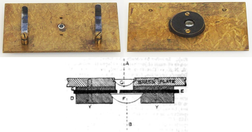
Figure 3. Lord S.G. Osborne’s “Diatomescope”, which was manufactured and sold by Ernest Hinton. Upper row: top and bottom views of a diatomescope. Lower row: Schematic cross-section, from Osborne’s description of his invention that was published in the November 21, 1884 issue of English Mechanic and World of Science: “The
optical arrangement consists of two piano convex lenses, F and G - F being
mounted in the metal disc D, so that its axis B is laterally excentrical in
relation to the axis A of the lens G. G is fixed in the centre of the brass
plate, which lies on the stage of the microscope. The plate C has a circular
central opening corresponding with the diameter of G, and appears to have been
inserted to regulate the distance between the lenses. The diaphragm plate E,
having a square opening 1/32 in., slides laterally in grooves on the disc D,
between the lenses, so that a pencil of light, striking the carved face of F
suitably, is refracted through the small aperture in the diaphragm plate E on
to the lens G, where it undergoes further refraction, and emerges from the
plane face at an angle of 60°, or more, in air, according to the obliquity of
the first incidence and the position of the sliding diaphragm. The slide to be
examined is placed fiat on the surface of the brass plate, with or without
immersion contact with the plane face of G, two spring-clips holding it in
place. D, C and the brass plate are connected together by the screws Y Y; these
screws, however, are not really inserted where figured, but in a diameter
passing through A, at right angles to the section shown.”
The editors of Hardwicke’s Science-Gossip quickly took note of Hinton’s production and sales of the Diatomescope, although they initially mistook it for Osborne’s hand-held device. This was immediately corrected, and expanded upon thusly: “The Diatomescope. — We are sorry that another and a quite different instrument was last month noticed under this name. The Diatomescope is constructed by Mr. E. Hinton, and our readers will find a full account of it in the ‘English Mechanic’ for August 22nd. It is a beautifully finished and ingenious adjunct to the microscope, being a kind of supplementary stage, which can be instantly placed on the ordinary stage. In the centre is inserted a powerful lens, so that the reflected light from the mirror beneath passes through it, and produces an exquisite side illumination of the objects viewed. In this manner the dots or striae of diatom, &c. are brought out with remarkable distinctness. Mr. Hinton has conferred a favour on microscopists by bringing out this cheap and effective auxiliary.” In addition, Henri van Heurck and George W. Royston-Piggott, two of the most prestigious diatomists of the period, wrote favorably in The English Mechanic on the Osborne-Hinton Diatomescope.

Figure 4.
An 1890 letter from Hinton to G. Kavanagh, Dublin, Ireland (courtesy of Richard Courtiour). The text reads,
“I have forwarded you per this afternoon post one of Lord Osborne’s Diatomescopes. I do not think that you will find any difficulty in using it. All that is really necessary is to have the lamp on the left hand of the microscope and about level with the mirror. If you find you do not get light enough move the little slot so as to bring the square aperture into the center of the upper lens but I think that you will find it about right as sent. Try it on the slide of Angulatum sent with it. This preparation is not sent as a specimen of mounting for it is by no means a good preparation, but it contains several different species of Pleurosigma and is therefore a very good test.
I should like to take this opportunity of calling your attention to my list of objectives, they are superb glasses and wonderfully cheap. Dr. Royston-Pigott, a very great authority on all matters relating to optics, writing to me about my cheap ¼, said ’you have no idea of the superlative quality of this ¼ for 2/-’ !!!!
I have no detailed list of my preparations for the microscope. I do not now publish one, but shall be most happy to forward a few dozen for your inspection if you would care to look through them, by Embryology slides do you mean Human or lower animals?
Yours truly
Ernest Hinton”
Royston-Piggott also praised Ernest Hinton’s microscope
slides, in particular, his mounts of diatoms and of butterfly scales.
Royston-Piggott was especially interested in microscopic resolution of the
tiniest features of such objects. As an example of Royston-Piggott’s enthusiasm
for Hinton’s diatom mounts, in 1886, he wrote, “A few days ago a friend brought an old, but very good, Powell and
Lealand 1-12th dry lens. We tried it on the Amphitetras ornata, and we could
both make out nothing at all; we were both astounded. The best P. and L. 1/8
showed everything superbly; this diatom, mounted by Hinton, can be strongly
recommended”. Many of his other articles on diatom studies specifically mentioned Hinton as the slide maker. In 1888, he wrote in the English Mechanic and World of Science,
“Mr. Hinton, Vorley-road, Holloway, has
sent me some most beautifully clear mounts, dry, of the following scales,
showing large villi: - Zygoena, Clipendube, athmenthae, coronillae, lavendulae,
transalpae, meliloti, trigonillae, medura.” The Wesley Naturalist wrote that same year, “Butterfly Dust and Villi: The use of improved lenses, new mounting
media, and careful illumination, is yielding excellent results even where we
should hardly have expected to make any further advances upon the stock of information
already obtained. In the English Mechanic for September 30th we have an
illustration of this statement which deserves a passing note. Dr. Royston-Piggott, M.A., has for some time past been
engaged in the study of the scales of insects, and by the use of castor oil has
been able to produce slides (which Mr. Hinton, of 12, Vorley Road, Upper Holloway, is mounting, in excellent style, by the
Doctor's instructions, for popular use), shewing that the scales of the Burnet
and other moths are clothed with ‘villi’ or minute appendages and beads, which
form admirable tests for high powers, while they further illustrate the
marvellous details of structure in humble forms of life.” In 1887, Hardwicke’s Science-Gossip reported that
“In the English Mechanic of September
30th is a remarkable article by Dr. Royston Pigott, on
‘Butterfly Dust, Villi, and Beads.’ The detection of villi in butterfly dust is
a new departure in microscopical work. Mr. Ernest Hinton, of
12 Vorley Road, Upper Holloway, has brought out a capital preparation of the
scales of a moth (Zygtena trigonilid) which illustrates the above paper on
villi in a remarkable manner. All microscopists will be interested in the
subject on account of its high importance.”
In a footnote to one of his 1886 papers on diatom resolution, Royston-Piggott wrote the following: “Hinton did nearly all the mounting for Wheeler, and his address is Vorley-road, Holloway. He lived with him 20 years, from 11 years of age, I have never seen Hinton's preparations surpassed.” Brian Bracegirdle noted this statement when he described Ernest Hinton in Microscopical Mounts and Mounters. Since Edmund Wheeler is known to have also employed his nephew, Frederick Enock, the statement cannot be completely true. According to statements he made to census takers, Edmund Wheeler’s primary profession was public lecturing, which undoubtedly cut into his time making slides and microscopes. However, Enock wrote (1904) that Wheeler produced a large number of slides himself, “After a six weeks’ ’Lecturing Spree’ as he called it, he would come home and in five minutes would be in his den, working with all his might, and so keep on till the midnight oil was burned”. In addition, Wheeler sold slides produced by other mounters, including (according to Enock), “the wonderful microscopic work of such men as the late Charles Fielding and his son Amos Topping (sic, Charles Morgan and Amos Topping), Rev. Thornton, Herr Maller (sic, Moller), and others who contributed to his enormous stock of objects”. The noted diatom mounter John Barnett also evidently distributed through Wheeler, and may have helped Hinton learn that art (see the biography of J. Barnett on this site). In addition, Wheeler stated in 1864 that his microscope slides were “prepared and mounted by himself and the other members of his own family, his sons and his daughters”. By 1871, however, one of Wheeler’s daughters had died, and the other daughter and his son were living in Brighton. Thus, Royston-Piggott’s comment was partially correct, with Edmund Wheeler making a fair number of slides, other mounters factoring through Wheeler, and Ernest Hinton and Fred Enock producing the lion’s share of the Wheeler output.
As noted above, Hinton began advertising his microscope
slides and Osborne’s Diatomescope as soon as the Wheeler business was closed.
Hinton also cleverly sent examples of his slides to editors of popular science
magazines such as Hardwicke’s
Science-Gossip. In 1885, that magazine wrote, “Type Slide of Blood. - Mr. Ernest Hinton also sends a slide, showing in
one mount the blood corpuscles of man, frog, bird, fish and snake, a very
compact and instructive method of showing the differences of type in the
several kinds of blood belonging to these different classes of vertebrate.”
In 1886, “New Slides - We have been
favoured with an admirably mounted set of slides, of Trichina spiralis, by Mr.
Ernest Hinton. No. 1 shows male and female; No. 2, the worm imbedded in the
muscle; No. 3, ditto (larva) dissected from muscle, and freed from surrounding
material; No. 4, Trichina in capsules; and No. 5, ditto calcined in the muscle.
All of them are of the highest use both to teacher and student”, and “New
Slides - We have received an admirably mounted and most instructive slide from
Mr. E. Hinton, 12 Vorley Road,
Upper Holloway, showing vertical section of an entire foetal mouse, in which are
all the principal organs and structures, eye, ear, brain, vertebrae, heart, lungs, kidney, spleen, intestines,
etc. It is impossible for a young biologist to work at a more profitable slide”.
In 1887, “New Slides - We have received
an admirably mounted and most interesting slide from Mr. Ernest Hinton, 12,
Vorley Road, Upper Holloway, of a desmid [Botryococcus Braunii), in conjugation”
and “Ova of the Hermit Crab - Mr. E. Hinton has sent us a most interesting slide of the ova of the hermit crab,
showing the filaments which unite the eggs together like a bunch of grapes, and
all of them to the abdominal segments of the female crab”. Hinton continued
to send free slides to Science-Gossip
through 1891, resulting in 2 to 4 free advertisements per year in that magazine.
The Pharmaceutical
Journal reported in 1897 that “Better
mounted objects than those prepared by Mr. Ernest Hinton are hardly
conceivable, assuming that the specimens he has submitted for examination
fairly represent his stock, as no doubt they do. The slides we have examined
are: Ovary of Tulip, T.S. (note: transverse section); Stem of Black Poplar,
T.S.; Stem of Scotch Fir, T.S.; Compound Spiral Vessels (Banana); Petiole of
Curled Dock, T.S.; Cystoliths In Leaf of Ficus. Most of the specimens are
double stained, the stains being clear and definite, thus assisting the student
rather than confusing him, as is sometimes the case. Special sets of a dozen or
more slides can be supplied to meet the requirements of pharmaceutical
students. The slides are specially prepared for educational purposes, and
students cannot do better than procure sets to illustrate typical structures.
For, whilst it is useless to expect to acquire a satisfactory practical
acquaintance with the details of vegetable histology by the mere study of
mounted objects not prepared by one's self, the value for reference purposes of
authentic and carefully prepared type specimens such as these is very great. In
addition to supplying educational and other slides, Mr. Hinton is prepared to
mount specimens to order from an investigator's own material. Price lists and
further particulars will be furnished on application to 12, Vorley Road, Upper
Holloway, London, N.”
Hinton is recorded as having taken advantage of other opportunities to show off his work. He exhibited “a good display of Histological, Botanical, and Pathological Slides” at the 1886 Annual Meeting of the British Medical Association, in Brighton. That meeting’s proceedings also noted that “Mr. Ernest Hinton (12, Vorley Road, Upper Holloway, N.) showed a Student’s Microscope, “the Diamond,” and a Case of Preparations for the Microscope.”
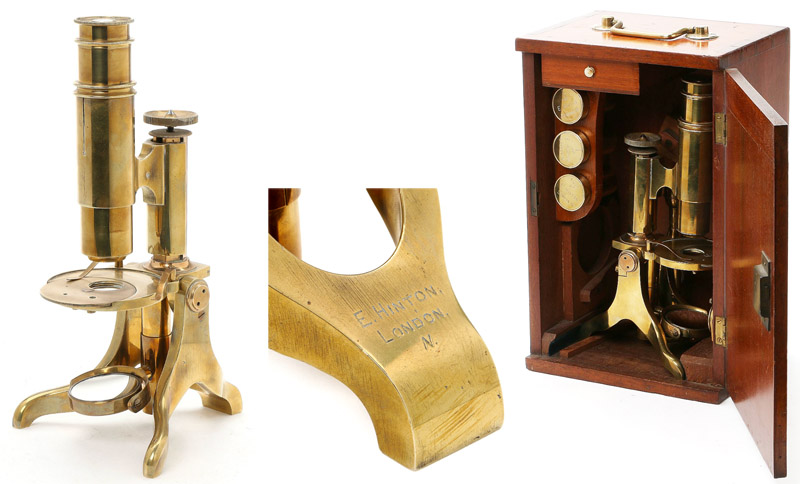
Figure 5.
A student-type microscope, marked by Ernest Hinton, which may be the “Diamond” model that he exhibited at the 1886 meeting of the British Medical Association. The letter from Hinton shown in Figure 4 states that he manufactured microscope lenses, suggesting that at least those components were made by him. Edmund Wheeler was a well-regarded maker of microscopes and telescopes, and it is reasonable to assume that Hinton would learn those skills from his master. Images from an internet auction site, used for nonprofit, education purposes.
An 1897 presentation to the Royal Microscopical Society was described thusly: “The President said he regretted there was nothing else upon the Agenda paper for the evening, excepting an exhibition of a number of very excellent specimens of injections and other objects by Mr. Ernest Hinton. These were shown under the Microscopes upon the tables, and would, no doubt, be inspected by the Fellows present with great pleasure and interest. He thought that the descriptive labels beside each instrument would render any further description unnecessary to those who saw the objects; but he felt that their thanks were due to Mr. Hinton for bringing them for exhibition. He therefore moved ‘ that a very hearty vote of thanks be given to Mr. Ernest Hinton, for affording this opportunity of examining these preparations’. The motion was then put from the chair, and carried unanimously. The meeting then resolved itself into a Conversazione, at which the following objects were exhibited: Mr. Ernest Hinton: - Feet of Toad (two preparations). Foot of Frog. Upper jaw of Toad. Under jaw of Toad. Surface of stomach of Toad injected with chrome. Ditto, ditto, injected with carmine. Surface of small intestine of Toad injected with chrome. Ditto, ditto, injected with carmine. Surface of skin of Toad from belly and back. Surface of human skin. Ova of Frog. Lung of Python injected with chrome. Ditto, ditto, injected with carmine. Large intestine of Python. Small intestine of Python injected with chrome. Ditto, ditto, injected with carmine. Small intestine of Grass Snake injected with chrome. Ditto, ditto, injected with carmine. Small intestine of Goat. Ditto of Lynx. Ditto of Emu. Large intestine of Emu. Leaf of Drosera rolundifolia showing captured insect. Heads of Chrygop relictus, Haemalopola pluvialit, Palloptera pulchella, Tryptera chrysanthemi, Tryptera reticulata, prepared without pressure, to show the eyes in their natural brilliant colours.” These would undoubtedly have been representative of Hinton’s productions at the time. It might also be expected that he carried duplicate slides to the demonstration, for sale to attendees. Note that Figure 1 of this essay (above) includes one such slide, labeled “Head of a Gall Fly from Chrysanthemum prepared to show the brilliant Eyes Tryptera Chrysanthemi”.
The Worshipful Company of Spectacle Makers (the guild of
opticians) put on a major show, The Exhibition of Optical, Mathematical, and
Scientific Instruments, in London during October, 1898. Ernest Hinton displayed
preparations for the microscope. A report on that exhibition also provides
insight into the “optician” trade of 1800s England: “At the time when the Company was granted its Royal Charter, in 1629,
spectacles were practically the only optical instruments dealt in, and the
comparatively few makers and sellers were all connected with the Guild. As
science progressed, and the demand for other optical and philosophical
instruments increased, the spectacle maker ceased to confine his trade to that
one article, and became the general optician, while from the fact that
spectacles are so generally in demand they became ordinary articles of
commerce, and their sale extended to trades totally unconnected with optics”.
Hinton continued to advertise and exhibit his products until near the time of his death. In 1905, he presented at the Annual Meeting
and Conversazione of the Selbourne Society.
Such was Hinton’s reputation as a slide preparer that his
works could be found throughout the world during his lifetime. On December 13,
1886, the Royal Society of New South Wales (Australia) exhibited “Several slides from Hinton (London) of those diatoms described by Dr. Royston Pigott in the ‘E. Mechanic’’.
Ernest Hinton joined the Quekett Microscopical Club on
January 18, 1895. That year, and probably during subsequent years, he donated
several of his slides to the Club’s cabinet. He remained a member of Quekett
until at least 1906. On October 16, 1896, a new microscope lamp produced by
Hinton was exhibited to the Club by William Goodwin, its inventor (Figures 6 and 7).
Several popular science magazines, including The English Mechanic and The
American Monthly Microscopical Journal picked up on the lamp: “This excellent lamp, which combines
portability with great efficiency, was designed and exhibited at the meeting of
the Quekett Microscopical Club, on the 16th of last October, by Mr. W. Goodwin,
a member of the club. The lamp which is nickel-plated, is 2 1/8 in. in diameter
6 ½ in. in height, and weighs about 3 oz. A glance at the figure shows that it
has a metal chimney with two openings: this makes it available for the
illumination of two microscopes at the same time. The burner takes a 1/2 in.
wick, which yields sufficient light for an amplification of 2,000 diameters
when a suitable condenser is used. The glasses are optically worked, one being
tinted steel blue, the other signal-green; if, however, untinted light is
desired, circles of thin cover glass may be used instead. These, if carefully
selected, will stand the heat of the flame without cracking. The lamp is so
small that it can easily be packed in the same case with the microscope, thus
dispensing with an extra box. The price of the lamp is about 12s., and it is
made by Mr. H. Hinton, 12 Vorley-road, Upper Holloway, N”.
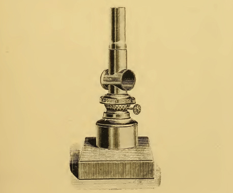
Figure 6.
Illustration of the Goodwin microscope lamp, as manufactured by Ernest Hinton.
The lamp has two openings to emit light, fitted with either a blue or green
glass. Adapted from the Journal of the Quekett Microscopical Club, 1896.
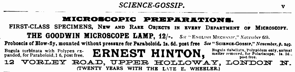
Figure 7.
An 1897 advertisement from Ernest Hinton, emphasizing the Goodwin microscope lamp.
Ernest’s wife, Clara, died November 18, 1889, from “exhaustion” following 5 years of
suffering from breast cancer. His mother, who had continued to live with
Ernest, died in 1893. During 1905 or 1906, Ernest moved from 12 Vorley Rd. to
11 Cornwallis Ave., Lower Edmonton, London. Ernest Hinton died there on October
10, 1909, from “perforation of intestines
due to cancer”. The Journal of the Quekett Microscopical Club wrote “The Secretary said he regretted to have to
announce the death of Mr. E. Hinton on October 10th under very
painful circumstances. Mr. Hinton
had been a frequent attendant at their meetings and was well known to many of
the members. An expression of regret and sympathy was sent from the meeting to
the relatives of the deceased”.
In summary, Ernest Hinton was a widely known and respected
commercial preparer of specimens for the microscope. He also manufactured and
sold microscope accessories, such as Goodwin’s lamp and Osborne’s diatomescope.
He may also have produced microscopes. Hinton worked for Edmund Wheeler for 20
years, and produced a large percentage of the slides sold under Wheeler’s name.
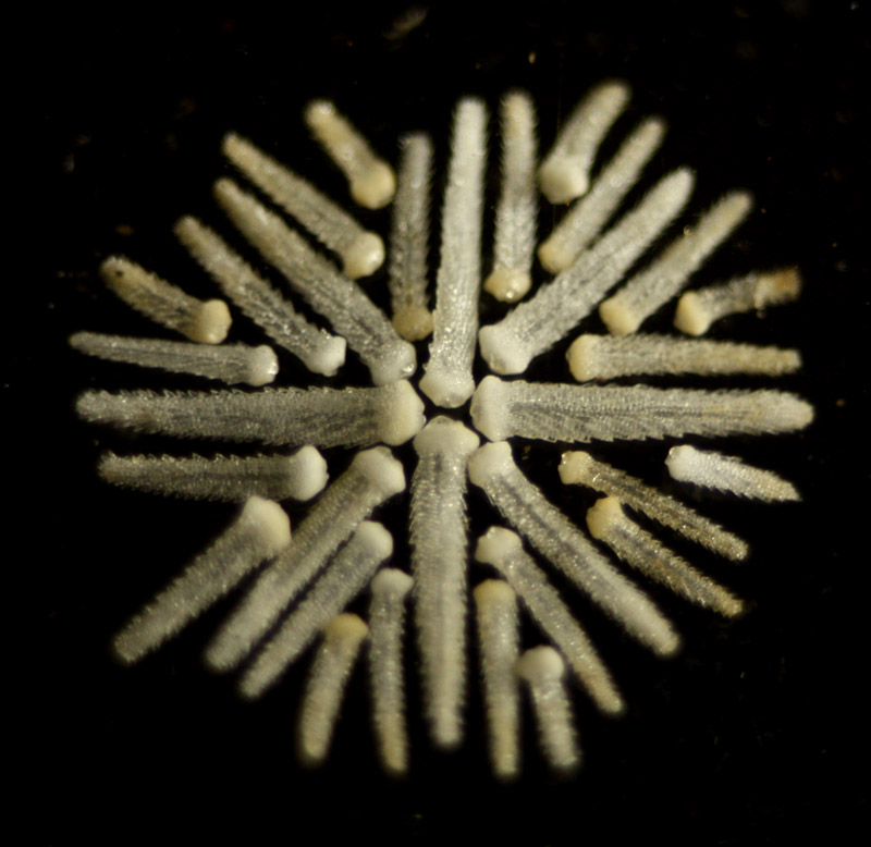
Figure 8. Detail of
arranged spines of starfish, by Ernest Hinton (see Figure 1, above).
Acknowledgements
Many thanks to Richard Courtiour for providing images of Ernest Hinton’s letter, and to Jurriaan de Groot for providing images of a diatomescope.
Resources
The American Monthly Microscopical Journal (1897) A new microscope lamp, Vol. 18, pages 128-129
Bracegirdle, Brian (1998) Microscopical Mounts and Mounters, Quekett Microscopical Club, London
The British Medical Journal (1886) Sept. 4, pages 453-462
Death record of Clara Hinton (1889)
Death record of Ernest Hinton (1909)
England census, birth, marriage and death records, accessed
through ancestry.co.uk
English Mechanic and World
of Science (1884) The Diatomescope, Vol 39, page 561
English Mechanic and World
of Science (1884) The Diatomescope, Vol 40, pages 18, 180-181, and
263-264
Giordano, Raymond V. (2006) Singular Beauty: Simple Microscopes from the Giordano Collection
Hardwicke’s Science-Gossip (1884)
The Diatomescope, Vol. 20, pages 257 and 276-277
Hardwicke’s Science-Gossip (1885)
Vol. 21, pages 42, 139 and ii
Hardwicke’s Science-Gossip (1886)
Vol. 22, pages 67 and 139
Hardwicke’s Science-Gossip (1887)
Vol. 23, pages 15-16, 187 and 259
Hardwicke’s Science-Gossip (1888)
Vol. 24, pages 163 and 234
Hardwicke’s Science-Gossip (1889)
Vol. 25, pages 43, 114, 168 and 211
Hardwicke’s Science-Gossip (1890)
Vol. 26, pages 41, 163 and 280
Hardwicke’s Science-Gossip (1892)
Vol. 28, page xvii
Hardwicke’s Science-Gossip (1897)
New Series, Vol. 3, page vi
Hodgson, J. Spence, Frederic Enock, and Wilfred Whitten
(1904) Edmund Wheeler, Reminiscences Of
Ackworth School, Ackworth Old Scholars' Association, report No. 23, pages
65-70.
John’s Family Tree, http://www.tree.me.uk/TNG/getperson.php?personID=I1079&tree=Tree
and related links, accessed June, 2010
Journal and Proceedings of
the Royal Society of New South Wales (1887) Vol 20, page 337
Journal of the Quekett Microscopical Club (1895) New Series, Vol. 6, pages 53 and 218, and List of members
Journal of the Quekett Microscopical Club (1896) A portable microscope lamp, New Series, Vol. 6,
pages 345
Journal of the Quekett Microscopical Club (1904) List of members, New Series, Vol. 9
Journal of the Quekett Microscopical Club (1906) List of members, New Series, Vol. 9
Journal of the Quekett Microscopical Club (1910) New Series, Vol. 11, page 33
Journal of the Royal
Microscopical Society (1897) pages 259-260
Marriage record of Joseph George Hinton and Sarah Maria
Blackmore (1845) Parish records of St. Paul, Canonbury, Islington
Nature (1897)
Diary of Societies, Vol. 56, page 48
Nature Notes: The Selbourne
Society’s Magazine (1905) The annual meeting and conversazione, Vol.
16, page 101
Pharmaceutical Journal (1897)
Microscopic objects for students, Vol. 57, page 246
Pharmaceutical Journal (1898)
Exhibition of optical, mathematical and scientific instruments, Vol. 57, page
246
Royston-Piggott, George W. (1886) Microscopical advances, English Mechanic and World of Science Vol
43, pages 203-204
Royston-Piggott, George W. (1886) Microscopical advances, English Mechanic and World of Science Vol. 43, page 402a
Royston-Piggott, George W. (1886) Microscopical advances -
XXXVI, Researches in high-power definition – attenuated lines, circles and dots, English Mechanic and World of Science Vol. 47, page 226
Royston-Piggott, George W. (1888) The villi and beading
discovered on butterfly and moth scales, The Journal of Microscopy and Natural Science, Vol. 7, pages 167-169
Stevenson, Brian, and Stanley Warren, The life and works of Edmund Wheeler, manuscript in progress
The Wesley Naturalist (1888) Butterfly dust and villi, Vol. 1, page 273