John Gibbons Hunt, 1826-1893
by Brian Stevenson
last updated March, 2015
J.G. Hunt was a physician and medical school professor in Philadelphia, USA, as well being an accomplished microscopist. He was reported to be an expert in mounting human and animal histological specimens, but few of those slides are known to have survived. His became an expert in botany, which he initially investigated with the techniques and stains he used for animal histology. He was an early practitioner of double-staining plant specimens. Despite the professional appearance of his slides, I have yet to find a reference to Hunt selling his preparations. A colleague wrote that Hunt “liberally distributed” his slides, suggesting that they were spread about by exchanges or gifts. He did exhibit mounts at the 1876 Centennial Exhibition in Philadelphia, but as an officer in the main local scientific society, his presence may have been to help show off the capability of Americans. Many of Hunt’s slides survive in excellent condition, and are macro- and microscopically very attractive.
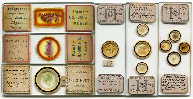
Figure 1.
Microscope slides prepared by J.G. Hunt, showing a variety of label types. The vast majority of known, surviving slides by Hunt are of botanical specimens.
High points of Hunt’s personal life are excerpted below, taken from memorials of the esteemed microscopist and physician. Following that are some details of Hunt’s microscopical work, and discussions of apparent results from his close relationship with microscope-maker Joseph Zentmayer.
Excerpts from J.G. Hunt’s obituary in The Proceedings of the American Microscopical Society, Harshberger’s Botanists of Philadelphia, and Kelly’s Cyclopedia of American Medical Biography:
“John Gibbons Hunt, physician and microscopist, was born at Darby, Pennsylvania, July 26, 1826, the son of Abram Gibbons Hunt, a farmer, and Massey Jones. He graduated M.D. at the University of Pennsylvania in 1850 with a thesis on ‘Histology of Muscular Tissue’. In 1858 he became a member of the Academy of Natural Sciences; in 1884 a fellow of the College of Physicians of Philadelphia; he was professor of histology and microscopy in the Women's Medical College, Philadelphia, 1872-1890. During the Civil War he was acting assistant surgeon U.S. Army in charge of Summit House Hospital, Philadelphia.
He married Anna Maria White, daughter of Joseph White of Philadelphia, in 1851. They had three daughters who were practicing physicians.
(He) was for a long time an intimate associate of Joseph Zentmayer in microscopy. Like Zentmayer, Dr. Hunt was not prolific in publication, although he contributed a number of short articles to the Cincinnati Medical News and some minor periodicals. As a manipulator of the microscope and preparer of objects he was unsurpassed, but he looked on this skill as only the means to an end - a knowledge of the objects themselves. Having made himself familiar with animal histology, he very early turned his attention to the anatomy of plants, of which he acquired an intimate knowledge. He was one of the very first to apply to plants the methods of staining that were in use for animal tissues, having begun before 1850; and in 1853 he first commenced double staining vegetable tissues by methods afterwards published by Dr. Beatty, of Baltimore, whose articles were widely quoted in the journals of this country and Europe.
It was as a teacher that Dr. Hunt exercised his greatest influence. A practicing physician for many years in Philadelphia, he still found time to give a great deal of attention to instructing medical students and others in the use and care of the microscope and in the preparation of microscopic slides and objects. He was Professor of Histology in the Women's Medical College for a number of years.
Founder of the Biological and Microscopical Section of the Academy of Natural Sciences, and Conservator from 1872 to 1880, Professor Hunt did much good work. He was the first professor appointed under the by-laws of the Academy to the chair of histology and microscopic technology, and although master of the most refined technique, he never received a large share of popular recognition on account of his native modesty and reserve”.
The Times (Philadelphia), reported on Tuesday, March 21, 1893, that, “Dr. J. Gibbons Hunt, the distinguished microscopist, is seriously ill at his home, at Lansdowne, Delaware county. Dr. Hunt was for many years in active medical practice in Philadelphia, acquiring a high reputation. Falling health compelled his retirement about ten years ago, since which time he has been living quietly at Lansdowne”. Hunt died at home on April 29, 1893
As quoted above, from the memorial notices, Hunt was a close colleague of microscope-maker Joseph Zentmayer. According to several contemporary sources, Hunt influenced the design of Zentmayer’s instruments. These appear to have been features that enhanced accuracy.
In 1859, the Proceeding of the American Philosophical Society reported, “Mr. Justice described a superior microscope stand made by Mr. Zentmayer, of Philadelphia, for and under the direction of Dr. Hunt, of Philadelphia, and embracing all the important late improvements. Dr. Le Conte added his testimony to the admirable skill of the mechanician, and supported it by refering to the stand lately made by him for Dr. Goddard, and to the stand now making for the Academy of Natural Science, which promises to be one of the best extant”. The microscope manufactured for the Academy was a “Grand American” model (Figure 2). It is possible that the instrument produced “under the direction of Dr. Hunt” was also a Grand American or something very similar.
In 1877, Zentmayer spoke of his microscopes to the Academy of Natural Sciences (Philadelphia). Referring to his “Centennial” model, he said, “About two years ago, in a conversation with Dr. J.G. Hunt, he pointed out the importance of having an arrangement for illuminating the object by an achromatic condenser in an oblique position. I explained to him how I would make a stand, in which this idea would be carried out in the most complete manner. The design and drawings were made soon after, but the instrument was not brought out, as I intended it for the Centennial Exhibition. Some of you have seen it before, and previous to bringing it to the exhibition, you recollect, it was shown at our meeting here, in April, 1876”. An obituary of Hunt stated, “He was an earnest advocate of the binocular, and when Zentmayer made for him one of the first ‘Centennial’ binocular microscopes, Mr. Wenham, the inventor of the binocular prism now in use, insisted on fitting the prism himself, and the tubes were sent to England for that purpose”. An example of a binocular “Centennial” microscope is shown in Figure 3.
Hunt wrote a lengthy report on the microscope exhibits at the 1876 Centennial Exposition, held in Philadelphia. His paper is reprinted in full at the end of this essay, but some points are worth highlighting:
Hunt raved that the Centennial model was, “the only microscope stand constructed on accurate scientific principles. All its optical and mechanical parts are built around one primary centre, which is the focal point of the instrument. When placed horizontally, in the position for drawing, the entire microscope revolves around a centre which lies perpendicularly under the optical focus. A graduated base gives facility for approximative measurement of the angular aperture of objectives. The top of the stage is elevated accurately to a level with this centre of rotation, and revolves concentrically around the focal point. The stage is accurately graduated, and is adjusted by screws which are turned only by screw-drivers, and when once centred is not easily disarranged by careless trifling with inviting milled-head screws. All the illuminating apparatus, including the mirror, turns around the same centre, remaining always in focus, and all degrees of oblique light from 1° to 90°, are read off at sight on a graduated index level with the stage. This obtaining and registering of obliquity was perfected two years ago, and similar facility is not found in any other microscope”. Zentmayer acknowledged Hunt’s suggestion of the accurately swinging substage and mirror, and Hunt’s excitement over the entire instrument being perfectly aligned suggests that he may also have been involved with implementing that feature.
He also credited Zentmayer for moving the fine adjustment action from the microscope tube and placing it on the limb (compare the former on the “Grand American”, Figure 2, with the latter on the “Centennial”, Figure 3, below). Hunt criticized Ross’ microscopes for lacking that feature, “the Ross instrument, in which the fine adjustment is removed from the upper and placed beneath the lower end of the body, is a great improvement over the old pattern. Greater accuracy of motion is secured along with improved appearance. The wart is placed under the nose instead of on it, that is all. Like most other English microscopes, the distance between the focal point and eye-piece is changed every time the fine adjustment is touched, and therefore the magnifying power is constantly altering, and is perceptible under highest powers”. In contrast, “The fine adjustment has been removed from the end of the body, the wart has been operated upon … by Zentmayer's process, and not a drop of blood was spilled. It has disappeared entirely. Still a peculiarly shaped, large milled-head graduated screw, which gives a comparatively rapid or extremely slow motion, moves a slide independent from the rack-motion, and focusses the entire optical body, thus always preserving the same relationship between the objective and eye-piece, an arrangement not found in any first-class English microscope”. This skilled microscopist’s enthusiasm leads one to suspect that Hunt had a hand in designing Zentmayer’s improvements.
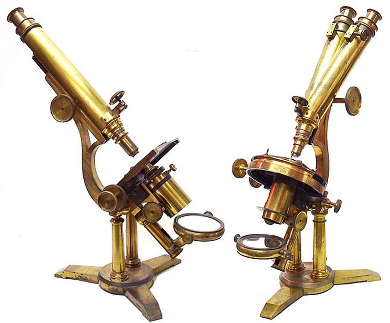
Figure 2.
Joseph Zentmayer’s “Grand American” microscopes, in monocular (left) and binocular (right) forms. Records suggest that he made a similar instrument for J.G. Hunt ca. 1859. Adapted, with permission, from http://www.antique-microscopes.com/photos/Zentmayer_Grand_American_microscope_monocular.htm
and
http://www.antique-microscopes.com/photos/Zentmayer_Grand_American_microscope_binocular.htm
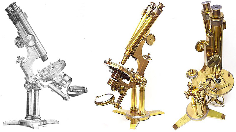
Figure 3.
Joseph Zentmayer’s ca. 1876 “Centennial” binocular microscope. Zentmayer made an early version for J.G. Hunt, who influenced the incorporation of the precisely-centered swinging substage and mirror. Judging from Hunt’s published views on microscopes, he may also have been involved with the design of Zentmayer’s fine-focusing mechanism. The engraving on the left is from the 1877 ‘Journal of the Franklin Institute’, and the photographs on the right are of a surviving microscope in the collection of Allan Wissner, adapted by permission from http://www.antique-microscopes.com/photos/Zentmayer_American_Centennial_Microscope.htm.
Hunt was a skilled preparer of human tissues for histological examination. In 1877, he co-produced a serial publication, “Microphotographs in Histology: Normal and Pathological”, along with Carl Seiler and Joseph G. Richardson (Figures 5 and 6). Reviews were generally positive, and supportive of the instructional values of the photographed preparations.
As noted above, J.G. Hunt exhibited microscope slides of botanical subjects at the 1876 Exposition. He was one of only four exhibitors to show substantial numbers of slides, so may have been present to fill up space. The others were William Walmsley (representing J.W. Queen of Philadelphia), George Beatty (of Baltimore), and Edmund Wheeler (of London). A review in The American Journal of Microscopy and Popular Science stated, “Amongst the objects shown were some very fine stained tissues by Dr. Hunt and Mr. Walmsley. The collection of the former (both animal and vegetable) is very large, and many of the specimens are of great value from the special features which they display”. In Henry Crouch’s report to the Quekett Microscopical Club, he stated, “I have the pleasure of submitting to your notice some very beautiful preparations of this description, mounted, and kindly presented to me by Dr. Hunt, of Philadelphia, which will be found to illustrate vegetable structure in a remarkable manner. As examples of mounting they are unsurpassed”.
R.H. Ward further praised Hunt’s slides, “The vegetable preparations, transparent and double stained, by (Hunt) are regarded with evident enthusiasm as remarkable illustrations of vegetable structure mounted in an unsurpassed manner. Dr. Hunt's exquisite transparent vegetable preparations can hardly be better appreciated anywhere than here … When first produced many years ago they were believed to be a large and important contribution to the progress of microscopy, but the methods worked out by Dr. Hunt were so unselfishly communicated, and the objects so liberally distributed and so largely studied and imitated, that they have long since become common property”.
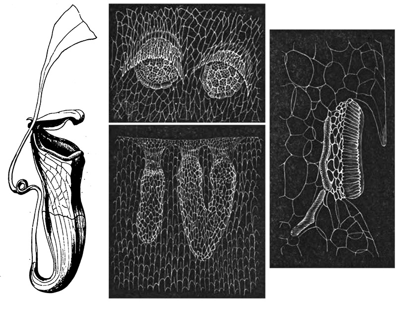
Figure 4.
Macro and microscopic drawing of the pitcher plant, Nepenthes distillatoria, by J.G. Hunt. Adapted from his 1870 paper on this plant.
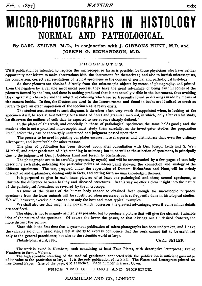
Figure 5.
An 1877 advertisement for Seiler, Hunt and Richardson’s “Micro-photographs in Histology: Normal and Pathological”. From ‘Nature’.
The full text of J.G. Hunt’s 1877 paper, “The Microscopes at the American Exhibition”, originally printed in the Cincinnati Medical Journal, and reprinted in the March, 1877 issue of The Monthly Microscopical Journal. The editor’s note from the latter journal is retained. As an aside, Henry Crouch attended the Exhibition, and apparently became friendly with Hunt, which may explain why Crouch was relatively unscathed by this critique.
“The Microscopes at the American Exhibition
By J. Gibbons Hunt, M.D., of Philadelphia, Pa.
[The following observations are remarkable from the bluntness, or rather sharpness, with which they are expressed. Still, if the reader will make the necessary allowances, they will not be found devoid of interest - Ed. 'M.M.J.']
After a great Exhibition, like the one recently held in our city, it may not be unprofitable to note some facts which have a bearing on that branch of human skill and science which is supposed to be cultivated in this section of the Academy, viz. microscopy.
Conscious incompetency would deter me from attempting a description of all the microscopical exhibits offered at our Centennial. I will ask you, therefore, to consider with me some subjects in which you and all workers with microscopes are interested, but which did not and could not find fitting expression in the reports of the eminent judges on that occasion. I take it for granted that we are at liberty to speak of the results of work, without embracing with admiration or neglecting with total indifference the workman, and this I propose to do from my own stand-point of observation, which is that of an interested observer of the field, rather than an active labourer therein.
Common courtesy leads me to speak first of the well-known foreign instruments which were displayed doubtless for the especial purpose of being looked at by all observers. Ornate show-cases have no essential connection with microscopy; they belong, in my opinion, to a distinct branch of mechanics. I shall not, therefore, entertain you with their description. Neither does needlessly massive brasswork necessarily give stability nor perfect motions to microscopes; therefore such specimens of brazen elephantiasis I will not further diagnose at this time.
The improved form of the Ross instrument in which the fine adjustment is removed from the upper and placed beneath the lower end of the body, is a great improvement over the old pattern. Greater accuracy of motion is secured along with improved appearance. The wart is placed under the nose instead of on it, that is all. Like most other English microscopes, the distance between the focal point and eye-piece is changed every time the fine adjustment is touched, and therefore the magnifying power is constantly altering, and is perceptible under highest powers. The new form is stronger and more steady than the old one, and less massive. The binocular prism is a fixture in the body, and does not change position while focussing. The Ross stage is still too thick, necessitating special and expensive apparatus for obliquest light. The finish of these instruments is good, but not the best, and the motions are smooth; but, I have reason to believe, had the hypercritical judgments of American microscopists been earlier known, that eminent firm would have displayed superior work to that we have seen.
Beck's large stand has more grace of form than any other foreign microscope; and, in excellence of finish, was superior to any other foreign instrument on exhibition. In my opinion the stage is mechanically defective. It has no adjustment for eccentric concentric rotation, and therefore seldom turns in the optical axis. Its mechanical arrangements for motion do not remain in order without frequent adjustment, and this results not from neglect of workmanship, but from defective design. Better abandon racks altogether in stage motions than spend time in adjusting bad ones. It is common experience, in this country, that foreign-made racks are not equal in smoothness of motion to those made at home.
The stands exhibited by Mr. Crouch displayed great excellence of workmanship, and this maker's aim has been to cheapen production without sacrificing commercial good work, and I think he has succeeded. His motions are made with more than ordinary foreign care, and his instruments therefore wear well. Crouch's best stands are supplied with the concentric adjustable stage, thus adopting Zentmayer's idea, introduced sixteen years ago. It is to be regretted that Mr. Crouch allowed his name to be connected with the introduction of the adjustable rotating stage, for it is exclusively an American invention.
The stands of Nachet are not elegant in design, neither is it the experience of workers here that they are conveniently adapted to all kinds of scientific work, nor do they continue in perfect condition after much use. I can remember the time when the American market was largely supplied with indifferent French microscopes, but, happily, that day is past.
Hartnack's instruments were not on exhibition, but previous experience has taught me they compare unfavourably with other reputed first-class instruments in workmanship and finish. After experience with American and English microscopes they are unsatisfactory in the extreme. Some German microscopists, and their imitators elsewhere, indulge the sickly sentimentality of lauding Hartnack's instruments as though they only were competent to do best work. In every respect, when compared with American and English first-class work, they are inferior. Clever working instruments in a restricted way they are, but they are not the best.
From Germany I have never seen first-class microscopical brasswork, and much of it has come under my notice. German microscopes are creations of deformity, and, speaking comparatively, are not instruments of precision at all. In the great struggle for the survival of the fittest, they will rapidly perish from sight, as rapidly as workers become instructed in such things.
American microscopes were in the minority at our Exhibition, if we estimate numbers alone. Not so if we consider beauty of design, workmanship, and originality of construction. Among such work, claiming to be first-class, Zentmayer's is pre-eminent. It has no superior anywhere. The stands he placed on exhibition were the best microscopical work there. In all his best stands the adjustable rotating concentric stage is used, and has been for sixteen years, long before any foreign maker conceived the idea.
The "American Centennial" stand, for the first time exhibited on that occasion, is worthy of special notice. It combines specialties of construction not found in any other instrument, and its mechanical finish is more perfect and displays superior workmanship to all others in the Exhibition or elsewhere. It is the only microscope stand constructed on accurate scientific principles. All its optical and mechanical parts are built around one primary centre, which is the focal point of the instrument. When placed horizontally, in the position for drawing, the entire microscope revolves around a centre which lies perpendicularly under the optical focus. A graduated base gives facility for approximative measurement of the angular aperture of objectives. The top of the stage is elevated accurately to a level with this centre of rotation, and revolves concentrically around the focal point. The stage is accurately graduated, and is adjusted by screws which are turned only by screw-drivers, and when once centred is not easily disarranged by careless trifling with inviting milled-head screws. All the illumina���������������������ting apparatus, including the mirror, turns around the same centre, remaining always in focus, and all degrees of oblique light from 1° to 90°, are read off at sight on a graduated index level with the stage. This obtaining and registering of obliquity was perfected two years ago, and similar facility is not found in any other microscope. Its scientific value is apparent*. By turning a large milled head screw a stage of extreme thinness, which likewise rotates concentrically, may be substituted for the longer one, and now your achromatic condenser and mirror may rise above the stage for illumination of opaque objects, and still the degrees are registered. The fine adjustment has been removed from the end of the body, the wart has been operated upon, not by Esmark's, but by Zentmayer's process, and not a drop of blood was spilled. It has disappeared entirely. Still a peculiarly shaped, large milled-head graduated screw, which gives a comparatively rapid or extremely slow motion, moves a slide independent from the rack-motion, and focusses the entire optical body, thus always preserving the same relationship between the objective and eye-piece, an arrangement not found in any first-class English microscope. The binocular prism is ground with equal skill and adjusted with more care than in most other instruments that have ever come under my examination; hence, both fields appear coincident, and do not resemble the longitudinal section of a cylinder, one side up, quarter way round depressed**. Here, then, we have a microscope of home production, but of surpassing precision, and which has taught the skilled English makers a useful lesson. If they propose to compete for the American market they must send hither better work. Thus far I have spoken chiefly of first-class microscopes, and only of those which have come under my notice.
* It is stated in the 'American Naturalist' for December, that a firm from Rochester, New York, "hinged the sub-stage bar at the level of the object," but the small stands exhibited by said firm at the opening of the Exhibition were not so made, neither had they any facility for registering obliquity. The firm in question did not grasp Zentmayer's idea at all, and hence can justly claim no priority of invention.
** The sub-stage is cut entirely through transversely, which gives unusual facilities for accessory illuminating apparatus.
The so-called student's stands are of equal importance, though less elaborate. All makers, foreign and domestic, furnish enough of these. Some are fit instruments for scientific work, very well adapted to the coarser observations in biology; but most of this class are not instruments of precision. It is a mistake to place in the hands of beginners bad tools to work with. Wherever a microscope is cheapened in cost by inferior workmanship, it is unfit for the student. It had better be in the hands of the expert who will eliminate its errors by his previous experience. Drop all mechanical luxuries in order to reduce expense, but give the best workmanship to the beginner. Much of this class of work sent here from abroad is so inferior that time would be wasted in speaking of it further.
In the construction of objectives great advances are to be noticed. On this subject my remarks will not be confined to lenses only which were on exhibition.
The patent system of Mr. Wenham, by which corrections are obtained by a single flint lens, was exhibited very fully before the judges. From the 1/5th to the 1/15th were on trial. They gave evidence of undeveloped microscopical potentialities of an advanced order, but their mounting and the mode of testing, justice compels me to say, were unsatisfactory. I therefore forbear judgment until I shall see more careful work.
Mr. Crouch's lenses were of the first grade. Those on exhibition and those seen since, without revealing any extraordinary optical qualities, are exceedingly fine in field and definition for their cost. Their corrections for achromatism resemble strikingly the Wenham lenses.
Beck's objectives form a series with which I am familiar, and they retain their character for many excellent optical properties. Without aiming at maximum angle, they are as nearly achromatic as lenses can be made. Their 4/10th is not inferior in performance to any other of equal power made, and, in use, is the most satisfactory lens of the series. But I must say these objectives - the adjustable ones - are not accurately mounted. The screw-collar jolts around from degree to degree in a way that forbids hope for the finest performance. The old plan of adjustment is retained, viz. of traversing the front combination, which must be comparatively defective. Lister's plan of adjustment and correction did well enough for twenty-five years ago, but modern microscopy demands a higher grade of work than that.
Fortunately, that demand is satisfied. The new 1/8th, so called by Powell and Lealand, brought into this non-achromatic world by what process of microscopical parturition we are not informed, ranks highest of all foreign objectives I have yet examined. Its corrections reveal a bluish-green light, and its definition marks an entire new era in English microscopy. It is difficult indeed to judge of this grade of lens because of our former defective experience. I cannot call its definition brilliant, but it is sharp and very accurate. On the margin of the field a good image is formed, which is generally not the case in lenses of extremest angle. The mechanical mounting and splendid finish of this truly grand objective should be a stimulus and admonition to other foreign makers to do likewise. It is useless to spend the time in such patient optical work as this lens demands if the mounting is defective. It is superbly mounted. Genius never clothes an angel of light in a beggar's garment. The American plan of traversing the back combination is adopted, and every expert knows its value.
From Germany I have seen nothing respectable. Several of Zeiss' objectives have come under notice recently. His lower powers which I have seen are unfit for use. His 1/25th fails in revealing details which our lower powers show better. The brasswork is specially inferior. Amplification is not definition. Power is necessary to transport mountains; definition and precision we demand in studying atoms. I do not see in Zeiss' objectives too much of Professor Abbe's mathematics, but I do see an absence of finger-skill which stamps them with a national characteristic. Mathematics never made an objective. Like theology, it says, this is the way, walk in it. It is the manner of walking in that way, in each case, which is the business. Yet these and similar grades use the lenses continually recommended to students. This is a serious mistake, and is the explanation of much misinterpretation in biological work. But these foreign lenses are cheap! For a dollar an optician will mount an uncorrected lens which will do as good work. Recall the results obtained by Swammerdam, who worked successfully at the anatomy of insects, and who discovered the values of the lymphatics in 1664. Of Leuwenhoek, who, with microscopes of his own make, better than some of��� which I am now speaking, and cheaper, discovered the organic muscular fibre cell, now attributed to Kölliker, and who described accurately the fibres of the crystalline lens of the eye. Of Malpighi and Grew, who first used the microscope in anatomy, and who made many discoveries in the structure of plants. Of Dr. Hook, and Baker, and Adams, and the earlier work of Ehrenberg. They observed with cheaper lenses and did better work than can be done with the glasses of which I am now speaking. Is anything cheap which misinterprets nature? Do you give the student in astronomical science, or in spectroscopy, or in surveying, or the chemist, or in any other branch of mechanics, bad tools to work with? Why should the biological student be specially degraded? Give him the best objectives. Cheapen their production as much as possible, but never at expense of their optical performance, because his function is to interpret, not only the genesis and structure of present organization; but equally, the vast and sacred mysteries of extinguished ages. True microscopy is the fertile branch of the great tree of aesthetics. Its revelations are the minute and beautiful things in nature, and when these are shown without optical distortion, we realize that splendour and grace are the common garments of all.
American objectives are not behind the best from abroad. I shall speak chiefly of those made by Mr. Tolles, because others of home product which I have seen have been disappointing. It is more difficult to judge of Mr. Tolles' work than of that of any other optician, because no two of his lenses that I have seen are alike; and that dissimilarity is evidently designed, and not accidental. Most surely his guide is not Lister, nor Amici, nor Abbe; but his genius is more comprehensive than all these combined. The true optician is he who can vary his formula at will to obtain other or finer results. To work by rule is mechanical, and may be taught an apprentice; it is never marked by progressive excellence. The power to direct your steps at will, while threading the labyrinth of optical construction, marks the master. That Mr. Tolles can do. In him are greater optical possibilities in the construction of lenses for the microscope than in any other maker, and my judgment is based solely on work. Still I have seen many lenses of his make which disappointed me greatly, because to gain some special point other qualities which I happened to value most were sacrificed. But when I detected, by larger experience, that all this was designed, and not accidental, my appreciation increased.
It is more amusing than instructive to hear learned professors define the limit of microscopical vision and the angle of aperture of objectives. They gravely tell us moreover that penetration and resolution are incompatible qualities in lenses. Possibly, in a degree, they may be so, but that degree is not yet a matter of professional experience. I can indicate objectives of Mr. Tolles' make of extreme angle, yet their penetration is so extraordinary, that they form the best lenses I know for best histological work by central light, showing details with a brilliancy which I never saw otherwise. A recent 1/10th which came into the world not by oblique presentation exclusively, is the highest standard to which I can refer. It is high commendation to compare any lens with Powell and Lealand's new 1/8th, but Mr. Tolles' last 1/10th is superior in most respects. Alike in power, the English lens has a remnant of London fog in its construction; the Boston one is brilliant and clear as crystal. Moreover, the Boston glass shows clearly structural details beyond the penetration of the English lens, without change of focus. Both are used wet or dry. The 1/8th has a separate front, the 1/10th is set for dry work by adjusting the screw-collar; this plan is more convenient than the separate front. A recent 1/5th, bearing the name of Spencer, from whom we naturally expect much, gave results not elsewhere obtained in lenses of that grade and cost - student's objective at $20.00 - but it was triumphantly under-corrected, and all ablaze with orange light.
I see cause to fear that micro-photography may, for a time, retard the best construction of lenses for histological work; especially that oblique micro-photography, whose best results are often only diffraction spectra, which leave it doubtful whether the lens or illumination was the chief factor in obtaining the result. Photography, at its best, gives only approximate representations of delicate structural details; and it is not yet proven that objectives so afflicted with strabismus are best for biological work.
Our best modern high-angle lenses have in them optical capacities not adequately developed by our present defective plans of illumination. Universal absence of absolute central light marks most microscopes, and accurate means of obtaining it, modified or concentrated at will, is a greater need at the present time, than further improvement in lenses. If we observe critically, all minutest details, as shown under most microscopes, are fringed with diffraction phenomena which can be removed often by simply improving the light. Even for coarser microscopical work attention to the light is universally neglected. Most instruments have no adequate provision whatever for accuracy of observation, hence misinterpretation is so common under the higher powers. The American microscopist has lenses, in common use, which will easily define Bacteria if our means of illumination shall be improved.
The results of microscopical work have interested our members on many occasions. Processes of demonstration, of comparatively recent origin, have given preparations of higher character than were attainable before. Our market is still too liberally supplied with foreign refuse material of this kind. Best work, in this department, is always kept at home. We import that which is unsaleable abroad. To this statement there are a few exceptions. In animal histology no one now hopes to see any foreign work worth having. In pathology, always more difficult of demonstration than normal tissues, we expect neither appreciation nor help from beyond the sea; yet it is not from talking members of pathological societies that we obtain best work. The Army Medical Museum, at Washington, has produced the finest pathological work, that is, work retaining most structural details, if not most neatly mounted that I have seen from other sources. In our Centennial there was nothing respectable from abroad in this department. Some of this imported stuff from Germany is abominable.
In demonstrating and mounting botanical subjects, this country is immeasurably in advance of all others. Some workers here offer preparations which are models of technological skill and of surpassing neatness. Every cell is revealed without dissection, and differentiated by double staining in most beautiful manner. But in this kind of work all structural details are not preserved. The cells are empty. None but the botanist will ever do best botanical microscopical work. If he knows not by previous study of the fresh tissues, what nature puts in them, he will not be successful in revealing them best in mounted preparations.
Biological science is not to be studied from microscopical slides, no more than from stuffed animals or dead shells in our museums. Botanists do not get best education by poring over mounted specimens, however beautiful they may be, no more than they do by daily browsing on the desiccated vegetation in herbaria or haystacks. We must go to the living for the best use of our instruments, and a knowledge thus obtained of structural detail is essential before any attempt should be made to preserve such details in mounted preparations. Some post-centennial work in botany, aiming at that result, has been exhibited before the Section, in which every cell showed the cell anatomy; the nucleolus, nucleus, protoplasmic contents, and cell-wall were all apparent at one view. Demonstration which falls short of this is unsatisfactory, because important morphological details are not brought out, and such work, like fossils in the rocks, belongs to a past era in microscopical technology”.
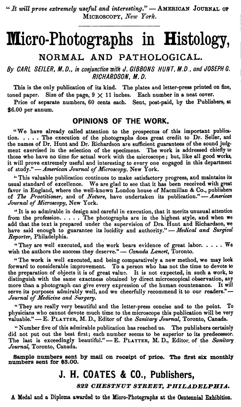
��Figure 6.
�1877 advertisement for “Micro-Photographs in Histology: Normal and Pathological”. From ‘The Boston Medical and Surgical Journal’.
Acknowledgement
Many thanks to Allan Wissner for permission to use images of his Zentmayer microscopes, adapted from http://www.antique-microscopes.com.
Resources
American Journal of Microscopy and Popular Science (1876) Microscopical conversazione (at the Centennial Exposition), Vol. 1, pages 131-132
Crouch, Henry (1876) On microscopy in the United States of America, Journal of the Quekett Microscopical Club, Vol. 4, pages 226-231
The Druggists' Circular and Chemical Gazette (1879) filler at end of page: “Dr. J.G. Hunt, of Philadelphia, one of the most experienced microscopists in the country, says that it is pure affectation or stupidity on the part of Americans to send to Europe for microscopes when they can get better ones at home for less money”, Vol. 23, page 102
English Mechanic and the World of Science (1877) Microscopes at the Centennial Exhibition, Vol. 25, pages 461-462
Harshberger, John W. (1899) John Gibbons Hunt, in The Botanists of Philadelphia and their Work, Harshberger, Philadelphia, pages 257-258
Hunt, J.G. (1869) Annual Address before the Natural History Club of Philadelphia (note: Hunt was then President of the Club)
Hunt, J.G. (1870) The structure of the pitcher plant, The American Naturalist, Vol. 3, pages 13-17
Hunt, J.G. (1877) The microscopes at the American Exhibition, Monthly Microscopical Journal, Vol. 18, pages 21-29
Hunt, J.G. (1880) Report on the annual exhibition of the Biological and Microscopical Section of the Academy of Natural Sciences, Philadelphia, held October 15, 1880, American Journal of Microscopy and Popular Science, Vol. 6, pages 20-23
Journal of the Franklin Institute (1877) Description of Zentmayer’s American Centennial microscope stand, Vol. 104, pages 52-53
Kelly, Howard A., and Walter L. Burrage (1920) Hunt, John Gibbons, A Cyclopedia of American Medical Biography, Vol. 1, Norman, Remington Company, Baltimore, pages 579-580
The Microscopical Bulletin and Science News (1893) Obituary and resolution by the Academy of Natural Science, of Philadelphia, Vol. 10, page 9
Proceedings of the American Philosophical Society (1859) Report of the meeting of June 17, 1859, Vol. 7, page 22
The Times (Philadelphia) (1893) note on J. G. Hunt’s illness, March 21, page 2
Ward, R.H. (1877) A foreign view of American microscopy, The American Naturalist, Vol. 11, pages 314-318
“W.H.S.” (1893) Obituary of J.G. Hunt, Proceedings of the American Microscopical Society, Vol. 14, pages 166-167
Wood, Horatio (1874) Collecting and preserving fresh water algae (includes a recipe for a damar-zinc oxide cement that was developed by J.G. Hunt), Journal of the Quekett Microscopical Club, Vol. 3, pages 192-197
Zentmayer, Joseph (1877) What I know about late improvements of the microscope, Journal of the Franklin Institute, Vol. 104, pages 49-51