Henry Pocklington, 1842 – 1913
by Brian Stevenson
last updated April, 2013
Microscope slides such as those illustrated in Figure 1
frequently create a lot of excitement at auction, and often sell for prices
well in excess of $100 each. The specimens are usually botanical, generally
leaves or fern fronds, although other subjects are occasionally found (Figure
2). The slides are well constructed, wrapped back and sides in a solid colored
paper, and the front covered with an off-the-shelf variety of gold-patterned cover paper. Specimen descriptions are nicely rendered in a distinctive hand, and are
written on plain paper below the cover paper, being visible through holes cut
in the covers. The holes cut through the covering papers to show the specimen
and the labels are imperfect, evidently cut by hand with a knife. That crude
feature points toward an amateur mounter, rather than a professional who would
have made use of cutting dies for clean, efficient cuts. Exact duplicates of
slides are not known, further suggesting that these slides originated from a
single private collection, rather from a commercial maker, who would
undoubtedly have made numerous identical slides of each specimen. The identity
of the maker of these attractive slides has long been a mystery for collectors.
Two slides have recently been discovered which suggest that the maker was Henry
Pocklington, a well-published authority in all aspects of microscopy.
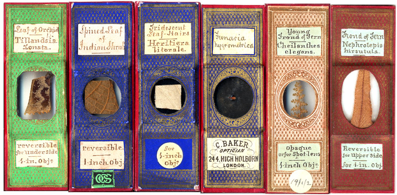
Figure 1. Examples of microscope slides that were probably
prepared by Henry Pocklington. He generally used the same pattern of
off-the-shelf cover paper, in colors that include green, blue, red and pink.
Although his slides are occasionally found with commercial dealers’ labels
attached (e.g. C. Baker, fourth from the left) – which might suggest a professional
preparer – it is important to remember that dealers such as Baker frequently
re-sold slides that they obtained from a variety of sources. The second slide
from the left has a label with the monogram “OCS” or “OSC” – the absence of
such stickers from the majority of these slides implies that it was applied by
a later owner. Bracegirdle’s Microscopical Mounts and Mounters illustrates two
slides by this maker, on Plate 40, one of which also carries an OCS/OSC
monogram, and the other a monogram of PCG, evidently another later owner.
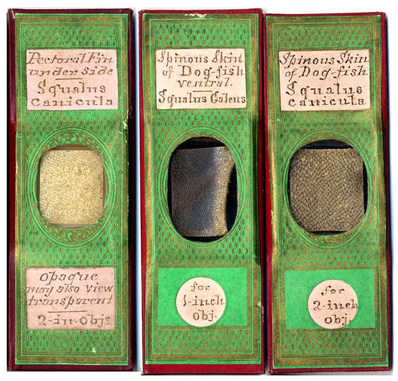
Figure 2. Three less common varieties of slides from this maker, being specimens of fish skins.
Two slides have come to light that bear important clues
about their most probable maker (Figure 3). Both contain leaf samples which are
labeled as being “Malvastrum mimroanum”.
This was a misspelling of the plant’s actual name, Malvastrum munroanum (now Sphaeralcea munroana). Both specimens came from
Idaho, USA, and one was recorded as having been supplied by Dr. J. Curtis of
the U.S. Geological Survey. Internet searches came up with only one record of
the plant’s misspelling, an 1872 English
Mechanic and World of Science article on “Leaves microscopically considered”, by “H.P.H.”:
“From the New World
has come a leaf ‘with stellate pubescence,’ interesting not only on account of
its hairs, but because of the place from whence my good friend Dr. Curtis
gathered it, and sent it from one extreme of this portion of cosmos to the
other. High up in the world in the new United States Park, at Idaho, with
‘literally thousands of geysers, and mud volcanoes round it,’ was this leaf
gathered, and there at least would one of its new friends like to go. My
friend's account of its habitat is interesting, and though out of place here,
should be incorporated, did space permit. The hairs on the surfaces of this
leaf (of Malvastrum mimroanum (?) gray) are radiate, the radii being of
considerable length and needle-shaped. Those on the lower surface are more
tufted, and somewhat resemble the hairs on the calyx of our homely English
mallows”.
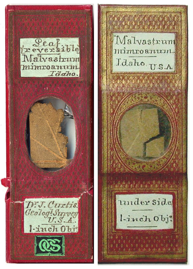
Figure 3. Two slides of “Malvastrum mimroanum”.
The misspelling, the attribution of Dr. Curtis as the specimen source, and the Idaho origin in both the publication and the slides all point to “H.P.H.” as the slides’ maker. Who was “H.P.H.”? Luck was again on our side, as a list is readily available on the web which gives the pen names used by frequent contributors to The English Mechanic and World of Science. “H.P.H.” was Henry Pocklington. His contributions were also often signed “H.P.,H.”, meaning “Henry Pocklington, of Hull”, that being the city in which he lived during the 1870s. Other Pocklington contributions were signed simply “H.P.”, and on a few occasions, with his full surname.
A third slide is known that also alludes to Pocklington
(Figure 4). This is an unpapered slide, with labels written in the same hand as
the above-described slides. It is a specimen of “sepiostaire” (cuttle fish bone) and includes a reference to an
article by Henry Pocklington on the subject (Figure 5). In light of the
above-described Malvastrum slides,
this slide was likely made by Pocklington, either for studies described in his
paper or for exchange with a colleague who was interested in the subject.
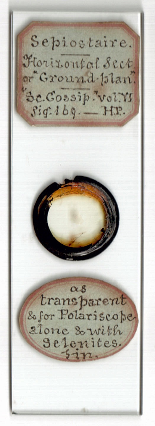
Figure 4. A microscope slide of “sepiostaire” (cuttle fish bone), citing an 1870 paper on the topic written by Henry Pocklington (see Figure 5).
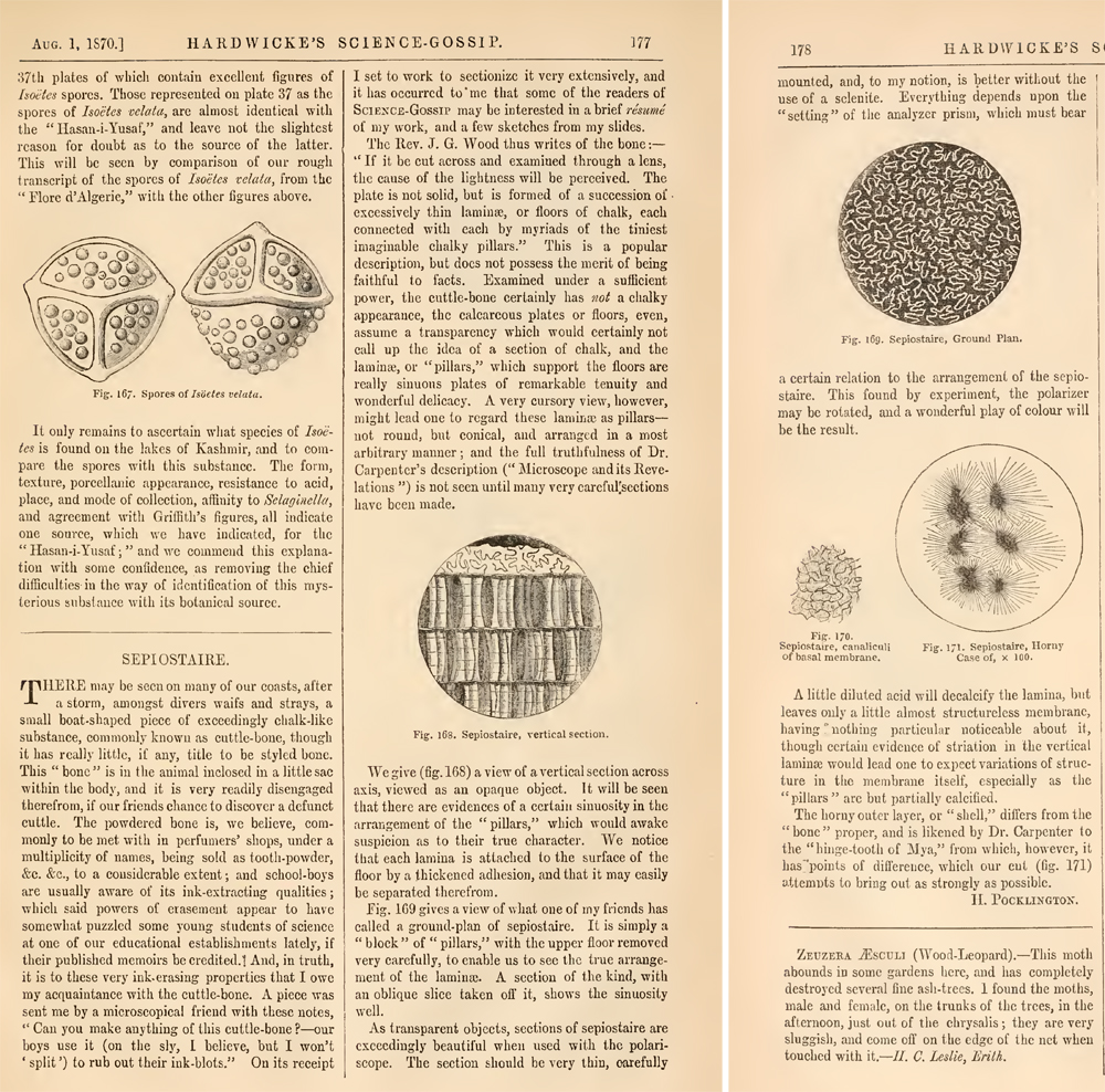
Figure 5. A paper on microscopical examination of sepiostaire,
by Henry Pocklington, that appeared during 1870 in Hardwicke’s Science-Gossip.
Muddying these conclusions is the existence of another slide
by the same maker, but with the name “Dr.
Herapath” on the label (Figure 6). William Bird Herapath was a medical
doctor and scientist. He is most famous for the discovery of “herapathite”, a
crystal of iodoquinine sulfate, which form the basis of Polaroid camera film.
Herapath was a man of his age, and explored a broad range of the sciences. In
1864, he described a new type of synapta (sea cucumber) acquired from Guernsey,
which he named Synapta gallieni vel
sarniensis (i.e. Synapta gallieni
or S. sarniensis, now known as Leptosynapta galliennii). The
microscope slide shown in Figure 6 is a specimen from that animal. I feel it
unlikely that Herapath made this slide, though. For one thing, no other known
slide produced by this maker is labeled “Herapath”
– if Herapath made all these slides, one would expect many of them to be
similarly labeled. Second, the honorific “Dr.”
would be excessive for someone labeling his own slides for his own collection,
even if he felt the need to write his name on the slides. Third, Herapath
primarily named the species “gallieni”,
after the man who collected the first specimens, so it is highly unlikely that
Herapath would have labeled a slide as only “Synapta sarniensis”. The strongest conclusion is that this slide
was actually made by someone else, probably Henry Pocklington, who either
attended or read about Herapath’s lecture, then made his own slide of the
synapta.
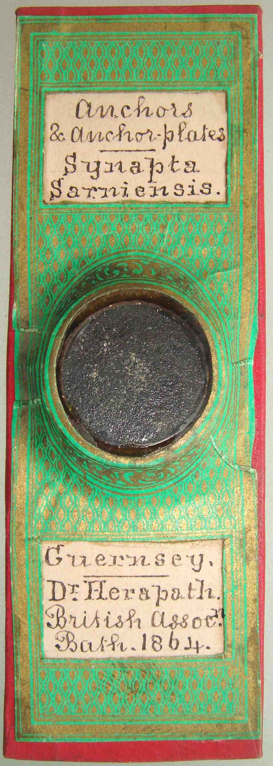
Figure 6. Anchors of the sea cucumber Synapta sarniensis. William B. Herapath actually named the species “Synapta gallieni vel sarniensis” (i.e. Synapta gallieni or S. sarniensis). Screen capture from an internet auction.
Henry Pocklington was born during late 1842 in Boston, Lincolnshire. He was the first child of Christopher and Eliza Pocklington, who had married the previous year. The 1841 census reported that Christopher was a “scrivener”, then living with his widowed mother, a “baker”, and his two brothers.
The 1851 census recorded Christopher, Eliza, son Henry and their three other children as living in Stilton, Huntingdonshire. Christopher’s mother lived with them. She was a “grocer general dealer”, and Christopher was a “shopman”. The family was doing moderately well financially, as they then employed an 18 year old girl as a live-in domestic servant.
Henry Pocklington initially planned to follow in his father’s and grandmother’s trade. The 1861 census recorded Henry as being a “grocers assistant”, living with and working for Edward Cooper in Cookham, Berkshire. He later entered the insurance business. In early 1869, Henry married Emma Janette Lilly, the daughter of an insurance manager. They had five children, two boys and three girls, although one girl died in infancy. Their eldest child, Henry Cabourn Pocklington, became renowned as a mathematician, physicist and astronomer, and was a Fellow of the Royal Society. Henry C. Pocklington’s 1953 obituary offered the following description of his father:
“Henry was self
taught; in his private life he interested himself in optics, astronomy,
electricity, mathematics and chemistry. This was at the time when the desire
for popular higher education was sweeping the country. Electrical instruments
were one of his hobbies; he used to make his children stand on small insulating
stools and hold the knob of some electrical apparatus, presumably of the
Wimshurst type. Miss Ida Pocklington (one of Henry’s daughters) recalled this
vividly; she remembered how her hair used to stand on end and how her scalp
used to tingle and feel uncomfortable for several days after such an
experience. Once H.C.P. received a severe shock and was thrown across the room.”
The oldest published record that I found which was probably written by Henry
Pocklington was a short note in Hardwicke’s
Science-Gossip, 1867, “A Stick
Without End - there may be seen in the churchyard at Shaftesbury, a somewhat
remarkable freak of nature. In the language of the foreman at the gas-works, it
is ‘a stick without an end’. A branch of a goodly elm has grown into, and
become part of another branch of the same tree, in such wise that it has become
really ‘a stick without an end’. - H. Pocklington”.
Exactly when Henry Pocklington became interested in
microscopy is not yet known to me. He was writing authoritatively in The English Mechanic and World of Science
by early 1870 (Volume 11 is the earliest to which I have access – if a reader
can provide information from prior issues, it will be greatly appreciated). The
tone of Pocklington’s contributions, plus the apparent familiarity readers had
with him, indicate that he was already a frequent contributor on microscopy.
His writings in 1870 indicate that he had been teaching classes in
microscopy for several years.
Of particular note, the July 8, 1870 issue included a
description of making microscope slides of cuttlebone or sepiostaire (see also
Figures 4 and 5, above). Other contributions by Pocklington in that volume
included a detailed description of the plant Anacharis, and notes on making slides of flower petals and
butterfly wings, where in England to find abundant diatom and foraminifera
specimens, microscopic investigation with polarised light, immersion lenses,
Wenham’s binocular, wood sections, plant physiology, mounting microscopical
objects with glycerine, arranging a microscope slide cabinet, the music of the
cricket, medical galvanism, and pre-Adamite man.
In general, Pocklington’s writings were both authoritative
and friendly, with frequent jokes and off-the-cuff remarks. This occasionally got
him into trouble. In an 1870 article on how to use the microscope and what
equipment was desirable, Pocklington remarked, “beware of opticians, unless you have a long purse: make what you want
when possible - that is nearly always”. The next week, he backpedaled,
writing, “As my caution to our readers
appears to have been misunderstood by some of the makers, I may, perhaps, be
allowed to say that nothing was further from my intention than to reflect upon
that most painstaking section; I merely wished to advise our readers to make as
much as possible and to buy as little as possible. It is only just to our
opticians to say that they are always willing to help the amateur to save his
pocket if he will trust himself to them”. That same year, he was also chastised
by professional microscope slide maker John Barnett about a comment that
appeared to rate Charles Topping’s work above that of Barnett. A series of
exchanges in The English Mechanic and
World of Science went as
follows:
“(Sept. 23, 1870) Can
any of your numerous microscopic readers give me any information respecting the
cleaning and preparing diatomaceous earths? I have been trying for a long time, and with very ill
success. I have this week been
examining some of Topping and Barnett’s preparations, and am disgusted with my
own efforts, - the last named gentleman’s slides being astonishingly
beautiful. If any of your
scientific readers can give me a hint as to the manipulation, I should be glad.
- F.G.
(Sept. 30, 1870) Perhaps
Dr. Carpenter's recipe will answer ‘F.G.'s’ purpose. It is to first wash the
earth several times in pure water, which should be well stirred and the
sediment allowed to subside several hours before the water is poured off. The
deposit is then to be treated in a flask or test-tube with hydrochloric acid
(muriatic acid or spirits of salts), and after the first effervescence is over
a gentle heat may be applied. As soon as the sediment has subsided the acid
should be poured off and another portion added, and this should be repeated so
long as any effect is produced. When hydrochloric acid ceases to act, strong
nitric acid should be substituted; and after the first effervescence is over, a
continuous heat of 200° Fahr. should be applied for some hours. When sufficient
time for subsidence has elapsed, the supernatant acid must be poured off and a
fresh portion added; the process being repeated so long as any effect is
produced. The sediment is then to be carefully washed until all trace of acid
is removed. This recipe will, of course, only apply to calcareous earths,
organic deposits, as guanos and the like. For siliceous deposits Professor
Baily's plan must be followed. This is to boil the deposit for a short time in
as weak an alkaline solution as possible (carbonate of soda as well as
anything). It must be borne in mind that this solution will act upon the
frustule as well, though not as much as upon the cementing matter. ‘F.G.’ must not expect to attain to Mr. Topping's pitch of excellence ‘all in a hurry’. - H.P.
(Oct 14, 1870) Mr. Barnett
complains that I have ignored him in my reply to the query of ‘F.G.’ a
fortnight since; need I say that I used, as probably ‘F.G,’ used, the two
names, Mr. Topping's name in a generic sense, and that nothing was further from
my intention than to exalt one professional preparer of objects at the expense
of another. As is well known to all microscopists, there are several gentlemen
of nearly equal merit, each of whom excels in some one department. Mr.
Barnett's preparations being certainly not inferior to those of Messrs.
Topping, Norman, or any other of half a score mounters, place him out of any
need for puffing. - H.P.
Perhaps to further make amends with Barnett, Pocklington
wrote in the November 4, 1870 edition: “put
a drop of the water containing the diatoms on a slip, and having allowed the
water to evaporate, may either mount them in water or dry, as he may elect. He
might do worse than arrange the diatoms nicely in a circle a la Mr. Barnett,
and then mount them in balsam. But as he probably will find this rather
difficult - I for one can't equal this maker's slides - he may be content if he
succeeds in keeping the diatoms separated, and does not float them out with the
wave of balsam”. The slide shown in Figure 7 may be an example of
Pocklington’s attempts to emulate Barnett.
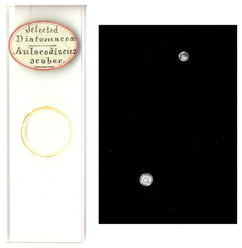
Figure 7. A microscope of selected diatoms, containing 3
randomly-placed circular diatoms. Only two of the diatoms are shown in the
photomicrograph, as the diatoms are so widely dispersed that they cannot all be
seen simultaneously with a 4x objective lens. This obviously amateur slide may
have been an attempt to copy the arranged diatom slides produced by
professional preparers such as John Barnett.
Pocklington also wrote, in December 1870, that “a pinch of most of the washing-powders used
in the kitchen or laundry department of our domiciles, dissolved in hot water
and allowed to re-crystallize in a thin stratum on a slip, and then mounted in
balsam forms a splendid object, preferred by many to even the gorgeous salicines
of Mr. Barnett. An immense number of delicately beautiful feathery stars are
seen on a dark ground when the object is viewed without a selenite”. It is
not known whether Barnett took that as a compliment or an attempt to deprive
him of business.
In 1871, the Pocklingtons lived in Sulcoates, Yorkshire.
Henry was then an inspector of his insurance company’s agents. From the
mid-1870s through the end of his Henry’s life, the Pocklingtons lived at
several addresses in the general area of Leeds, Yorkshire.
In 1872, Pocklington wrote the above-described article on
how to examine plant leaves under the microscope, which included the description
of “Malvastrum mimroanum” (Figure 4).
Also in 1872, Pocklington wrote a series of article for the Pharmaceutical Journal, on “the microscope in pharmacy”. He
contended that a pharmacist should be able to examine his wares for purity
under the microscope. He also advocated for pharmacist education to include
microscopic examination of beneficial plants and other objects. His papers
specifically described examination of beeswax for adulterants and
identification of samples of “Conii
Fructus” (hemlock fruit) contaminated with a poisonous plant. In 1873,
Pocklington lectured on “sugar and its
adulterations”. His son’s obituary commented on Henry Pocklington’s skill
and enthusiasm for ensuring product purity, “Henry became quite an expert at analyzing substances to be tested for
adulteration; it seems that samples of sugar had frequently to be investigated.
Chemical and optical methods (involving the use of a polarimeter) were used. He
became so expert that his friends among the city analysts often used to ask him
to visit their laboratories and check their work.”
I located only one published offer from Henry Pocklington to
exchange microscopical objects with other enthusiasts, in an 1873 issue of Hardwicke’s Science-Gossip, “Aecidium are offered in exchange for equally
rare fungi, good botanical or polariscope objects. - Henry Pocklington, 12,
Margaret Street, Hull”.
In January, 1875, Henry Pocklington was elected to be a
Fellow of the Royal Microscopical Society. In September, 1877, Pocklington was
voted into the Quekett Microscopical Club. He was also a member of the Leeds
Naturalists’ Club and Scientific Association from 1874 onward, serving as
president in 1878. He joined the Leeds Geological Association in 1874. In 1881,
he was elected to the British Association for the Advancement of Science, and
in 1898, joined the Royal Society of Arts.
Leading Insurance Men of the British Empire (1892) wrote, “Pocklington, Henry, the Leeds Manager of the Commercial Union
Assurance Company, entered upon his career of a District Manager in the year
1869, in the service of the Britannia Fire Association, whom he represented
first at Exeter and Hull, and subsequently at Leeds, having supervision of the
counties of Yorkshire, Durham, Northumberland, and Lincolnshire. He at the same
time acted for other offices, in the capacity of Surveyor and Assessor of
Losses. He entered upon his present duties as District Manager for the
Commercial Union Assurance Company in January of 1878, when that Company opened
their Yorkshire Branch, and being in want of a good Insurance man to supervise
its business, were fortunate enough to obtain Mr. Pocklington's services. Mr.
Pocklington was at one time a frequent contributor to the scientific press. He
is very strongly in favour of the thorough practical education in their
business of young Insurance men, and devotes much of his spare time to the
furtherance of this object. He is a Fellow of the Royal Microscopical Society of many years' standing, a Member of the Yorkshire Geological
and Polytechnical Society, of the Society of Chemical Industries, and of
several other scientific and literary societies”.
For the year 1907, Henry served as President of the
Insurance Institute of Yorkshire. The 1908 Proceedings
of the institute included a copy of the President’s year-end address, and a
picture of Henry Pocklington, which is reproduced below as Figure 8.
Pocklington was presented a gold watch on December 31, 1907 for his 30 years’
service as Leeds District Manager of the Commercial Union Assurance Company.
Henry’s wife, Emma Janette, died in 1908, at the age of 65.
In 1913, the Journal of the Royal Society of Arts reported, “Mr. Henry Pocklington, who died at Leeds on
the 13th inst., was for thirty years local manager of the Commercial Union
Insurance Company. He joined the Royal Society of Arts in 1898, and he was also
a member of the Society of Chemical Industry, the Royal Microscopical
Societies, and various other scientific bodies. At one time he conducted some
researches for the Royal Pharmaceutical Society into the adulteration of food
and its detection by microscopical methods. He was a keen photographer, and
devoted much attention to microphotographs. Deeply interested in education, he
took a prominent part in forming the Mechanics' Institutes of the North of
England, and many of the popular science classes in Leeds, Hull and Bradford
owe their origin to him”. He was then 70 years old.
________________________
During 1870, Henry Pocklington wrote a series of article on “the microscope – how to chose it and how to
use it”, for The English Mechanic and
World of Science. Excerpts are reprinted below, both as an example of Pocklington’s
proficiency with the microscope and for general interest of the historical and modern microscopist:
“As the optical
principles of the microscope are essentially the same as those of the
telescope, and have been so frequently treated upon in the pages of this
journal, it would be unwise to occupy further space than by saying that a
microscope is simply a means by which the eye of the observer is removed
further from the object observed, whilst the telescope carries the eye nearer
to the object in its field of view. The microscope may be either ‘simple’ or
‘compound’. The former class is commonly represented by the single lens of the
botanist, but a simple microscope may consist of a combination of several
lenses, arranged so as to act as a single one. Of the latter class are
‘doublets’ and ‘triplets’. The compound microscope necessarily consists of at
least two lenses, one of which is called the eye-glass, the other the
object-glass, the two or more lenses being usually connected by a tube of
metal. By the use of the compound microscope we obtain a much greater
amplification than is possible with a simple lens, inasmuch as the eye receives
a magnification of the image formed by the object-glass, and not the image
itself. A microscope of this land may be very easily made by any one possessing
the least mechanical ingenuity, but when made will be useless. Every object
viewed by its aid will be seen to be surrounded by a ‘beautiful’ coloured
fringe, and to be terribly distorted. These several defects, known as chromatic
and spherical aberrations, were for a long time insuperable obstacles in the
way of microscopic progress; but, thanks to ceaseless effort on the part of the
fathers of our science, we may now say that our instrument is about as perfect
as can be desired; and that the tale it tells is in the main true and faithful.
It is the compound achromatic microscope of which we intend to speak in these
papers. To this achromatic microscope there are two essential parts - the
mechanical and the optical - i.e., the stand and the lenses.
We now come naturally
enough to the enquiry - What constitutes a good microscope? Certain things must
be essential. What are they? 1st, as regards the stand. This must be solid,
heavy, so that it may be free from vibration, and well balanced. It must be
capable of being placed in either a vertical, an inclined, or a horizontal
position, and of remaining there without being clamped. The stage should be
sufficiently large to admit either edge of a glass slide, 2" in diameter,
to be brought under the object-glass. The aperture in the stage should not be
less than 1 1/2" or 2" in diameter, and the stage should be thin to
allow the oblique pencil to be thrown by the mirror upon any object on the
stage. The stage may be either simple or mechanical. If the former be chosen,
either the ‘magnetic stage’, the ‘lever stage’, or the ‘concentric rotating’
stage will be found useful. The plan adopted by Messrs. Beck is useful and
exceedingly simple, but with high powers is slightly tantalizing, as the focus
is disturbed by every movement of the stage, which is merely a thin plate of
metal held down by a double spring, the pressure of which may be regulated by a
screw (in practice it is advisable to screw this down tightly, as otherwise it
has an awkward knack of flying in one's face, to the serious detriment of one's
nerve, and possibly of the object), and this plate is doubled under the stage
on one side, so as to be grasped by the thumb and forefinger of the right hand.
This stage is extremely useful to the working microscopist, and after some
years of use we are disposed to speak very highly of it. There is nothing to
get out of order, and practised fingers will perform all needful movements
quite as delicately as would be possible with the most elaborate ‘mechanical’
stage. Below the stage should be fixed a diaphragm, which should be furnished
with a series of holes, in order that a variety of apertures may be available,
and the whole arrangement should be capable of being easily turned aside. Mr. Collins's ‘graduated diaphragm’ is perhaps the best possible. The mirror should be full
sized and double (concave on one side, plane on the other), and should be
capable of movement in all directions, as well as of adjustment nearer to or
further from the stage. It is convenient if the mirror be carried by a jointed
arm, as a more oblique illumination may be thus obtained.
Every microscope should
have a coarse and fine adjustment. The former may be obtained by a
rack-and-pinion movement, by a chain and pulleys, or by a watch-spring band.
The chain movement is peculiarly smooth and easy, and in practised hands
entirely obviates the necessity for a fine movement. This latter is usually
obtained by the action of a finely cut screw on a lever. The screw may be graduated
so that the distance through which the object-glass passes may be measured and
the thickness of an object approximately obtained. The milled heads of all
these adjustments should be so placed as to be conveniently accessible, and
they must work smoothly or they are utterly worthless.
A student's microscope is
usually furnished with two eyepieces, called A and B, being, as nearly as
possible, in the following ratios, 1, 2 ; and with two object-glasses - or, to
use henceforward the correct technical term - objectives of 1" and
1/2" focus respectively (i.e. these lenses have the same magnifying power
as simple lenses of those foci). The range of these powers is about as follows:
55, 90, 210, 350 diameters. These should be accurately centered and be perfectly
corrected. Means of estimating the quality of these lenses shall be given
later. A stage condenser and a stand, or bull's-eye condenser, for opaque
illumination, will complete the instrument.
The next question that
arises is, where shall we go for our instrument? Mr. E. Ray Lankester has
lately written in praise of foreign instruments; but we do not see what there
is to be gained by going abroad for that which may be obtained better at home.
English makers will beat the world for quality, and now - thanks to the Society
of Arts and some of our more enterprising manufacturers, - a really good
English instrument may be obtained at about the same price as the continental
ones. There is hardly any comparison between the convenience of the two classes
of instruments. These remarks apply only to the stands.
In lenses, the Germans
surpass us by far, Price being taken into account, although within the last
year our English makers have contrived to turn out very decent lenses at less
than half the prices formerly charged. We need only instance Crouch's or
Swift's 1/2" and Mr. Wheeler's 1/4". We will, therefore, look at home
for our stand. Where all are equally good it is a difficult (not to say an
invidious) task to instance the best. Those who wish a better-finished class of
workmanship may either select their higher priced stands, or look over the
catalogues of half a hundred makers and make their choice. The better plan is
to select a good stand, capable of being increased as funds are available, and
to add objectives and accessories from time to time. Such a stand will cost
about £10 or £15 with two eyepieces. The price of objectives will vary with
different makers. A fair English inch may be purchased for £2 10s., and a good
quarter for about £3. German lenses (Grundlach, of Berlin) of these foci will
not cost more than 17s. 6d. and 21s. respectively, and are about equal in
quality. Of course the first-class lenses of our best makers are unequalled by
those of any continental maker; but Messrs. Beck's first-class 1/4" costs
£5 5s. - a sum as large as many can afford to spend on the whole affair. To
such we commend the German lenses.
It will have been seen in
what we have said that the essentials of a stand are steadiness in all
positions of the body, ample stage room, and proper adaptation for the
reception of extra apparatus. The appearance of an instrument is of secondary
importance. To test the steadiness of the instrument use the 1/4" power,
focus carefully, and get some one to walk sharply round the room whilst you
observe an object. If there be excessive tremor, reject the instrument at once.
Next, try the adjustments, and see that they work smoothly and without ‘loss of
time’ - i.e., that they ‘answer"’ promptly to the slightest movement of
the milled heads in either direction. Use the 1" and 1/4" objectives,
and also a 3", and see that a small object remains truly in the centre of
the field of each power, and that there is no ‘twist’ or sideway movement on
altering the focus. So far for the mechanical portion of the instrument. We
will add that a short-bodied microscope having a draw-tube for elongation when
increased power is desired, is much to be recommended, on the ground that it is
far easier to work with. The corrections of the lenses must be carefully tested,
and unless the tyro go to a good and well-reputed maker, we would advise him to
get some experienced friend to select his lenses for him. The lenses of even
the best makers vary considerably, so that it is possible for an experienced
man to select a far better lens than might fall to his lot. The power of
1" should not be less than 30 diameters with the A eyepiece. It should
give a large, clear field, free from colour, and with a clean, sharp, circular
margin. For chromatic aberration the severest test is said to be a radial
section of fir. The glandular markings in this should be well shown, and be
free from colour with the C eyepiece. For flatness of field the section of an
Echinus spine is useful. For definition the pollen of mallow. For 1/4" a
good test for definition is the scale of the Podura or the frustules of
Pleurosigma hippocampus. The markings on either of these should be clearly
resolved. Dr. Carpenter specially recommends Mr. Lealand's preparations of
muscular fibre as giving a fair test for lenses of from 4-10th" to
1-5th" focus. Every objective should be tested with a series of eyepieces,
as a glass will often perform well with a shallow eyepiece, when a deeper one
will render manifest the most atrocious defects.
Having selected our instrument,
we will proceed to use it. Before us lie slides of Echinus, of Foraminifera
(mounted as opaques), of Diatoms, and the eye of a fly. Having taken our
instrument out of its case and put it in order (the maker of each instrument
will put the purchaser in the way of doing this), we will select a table having
a good light. If in the daytime, we will avoid direct sunlight as having too
much glare, and select a position in which we can receive light from a white
cloud. The microscope should be placed in an inclined position, and the mirror
adjusted so as to throw, an equable light upon the slide, neither too intense,
or too much the reverse. Careful use of the diaphragm apertures and focussing
of the mirror will give us any variety. We now take our slide of Echinus, and
having an inch objective on, place the slide on the stage and in the field. We
run down the rack motion until the objective is brought within 1/4" of the
slide, and then, with our eye to the eyepiece, focus back until we obtain a
clear definition. Having turned aside the diaphragm, we proceed to tilt the
minor into different positions, in order to get various degrees of obliquity of
the illuminating pencil. We will substitute a slide of Foraminifera for the
Echinus spine, and proceed to examine it as an opaque. Having closed the
aperture of the diaphragm, we throw a good light on the object by means of the
bull's eye, varying its angle of obliquity until we gain the best effect. The
working of the 1/4" is essentially the same, but unless we have the aid of
accessories, ‘transparent’ objects alone can be used with it, and greater care
must be paid to the focussing, &c, of the mirror. We cannot urge too
strongly upon our readers the importance of paying special attention to this
vital, but, to the beginner, seemingly unimportant, matter of illumination, as
truthful interpretation almost entirely depends upon it. The best possible
light for microscoping is daylight from a white cloud, but as most of our
readers (the author amongst the number) are compelled to work almost solely by
night, it is encouraging to know that good results may be obtained from the use
of a candle, even if it be protected by a glass shade, and it be not more than
10in. or 15in. from the instrument. The author has used for some years a small
paraffine lamp, costing at the outset about eighteen pence, and has found it to
answer every purpose, and to be most convenient, inasmuch as it permits a vast
variety of dodges in illumination to be tried with little trouble. And here let
us whisper to the readers of the Mechanic, ‘beware of opticians, unless you
have a long purse: make what you want when possible - that is nearly always’.
We have now, we think, gone
through our A B C. We have seen what our tool is - what are its essentials, and
how to put it through its A B C. But that is not learning how to use the
microscope. We have but learned to make a plaything of it, or at best to look
at bought slides. Again, we have but learned the use of a very simple and
unadorned compound microscope. We will, accordingly, if our readers be not
a-weary, glance at the use of a few accessories, and then try to learn how to
use the microscope whether in its simplest or most complete form.
We do not propose any rigid
order of sequence in our further notes upon the microscope, but we will
endeavour to take the several pieces of apparatus as they appear to stand in
the order of desirability. To this end we will allow ourselves considerable
latitude in our interpretation of the word accessory, and include therein all
apparatus not supplied with an ordinary student's microscope. And the question
we will set ourselves to answer shall be, What extra apparatus, and in what
order, would you recommend to a student ? I think we may place the polariscope
in the first place, partly because whether we do or no the student is tolerably
sure to do so, and partly because in proper hands it is an invaluable
instrument of research. In the form most commonly applied to the microscope the
polariscope consists of two parts - the polarizer and the analyzer. These are
precisely similar in construction and may (provided their fittings admit) be
interchanged at pleasure. The polarizer and also the analyzer may consist of a
rhomb of Iceland spar, or a thin plate of tourmaline or of herepathite
(sometimes called artificial tourmaline). On account of its cheapness and
freedom from colour, as well as its greater freedom from danger of accidental
damage, the Iceland spar is generally used, and in the form known as Nicol's
prism. Instead of the spar as a polarizer thin plates of glass may be used, but
as they do not permit of rotation readily their disadvantages are great. The
polarizer fits into the stage from beneath, and it is well if a bayonet catch
or some simple appliance be arranged to hold it firmly in position. The prisms
are fitted into a collar, which rotates easily by a milled head, unless the
stage be an elaborate mechanical affair, when special arrangements are made to
receive the prisms. The analyzer may be fitted either immediately above the
objective, in which position there is some loss of definition, or above the
eye-piece, in which case there is a considerable loss of field. In general work
the former position is the best. In any case the prisms should be made capable of
rotation with exact centering. If it be desired to carry the analyzer above the
eye-piece either tourmaline or herepathite should in all cases be preferred to
Iceland spar. The price of a good polariscope and fittings adapted for a
student's microscope varies from 30s. upwards. A selenite of some kind is
almost a necessary accompaniment of a polariscope. Sometimes a thin film (to
give red, yellow, or blue, according to fancy) is fixed in a collar and made to
rotate with the polarizer. But this is a barbarous arrangement. In all cases
the film of selenite should be mounted on a glass or in a brass slide, and some
means adopted by which it may be removed from the field at pleasure without
disturbing the object. It is also convenient to have three films giving
different colours set in the same frame, their axes of polarization being in
the same plane, that a series may be tried without trouble. A very efficient
selenite stage has been described in a recent number of this journal, the
price, however, is past a joke to most.
Having obtained the
polariscope, the student, if he be a worker and use his 1/4" much, will
find that he needs a steady and pure light such as is thrown upon the field by
what is known as the achromatic condenser. This is optically little other than
an objective of 1/2" or 1/4" focus made capable of approximation to
the lower surface of the slide under view. It is a somewhat expensive piece of
apparatus, costing from 30s. upwards. Mr. Brooke recommends a Kellner eye-piece
in the place of the usual short focus objective. In any case the student may
remember that he can always use his 1/2" objective as a condenser for his
1/4", and so on. In this case he need only purchase fittings which need
not be very costly. Mr. Collins has, within the last few years, introduced a
new condenser, which he calls the Webster. From what we hear of it it seems a
most efficient affair, especially in connection with his adjusting diaphragm.
Our student will now
probably seek to increase his ‘power’. He can do this by the addition of a
draw-tube to his instrument at the cost of a few shillings, but unless his
lenses are very good this plan is not to be recommended. The drawtube in
connection with an erector (for rectifying the inversion produced in the
apparent position and movement of objects when they are viewed through a
compound mirror) is, however, most useful, as it enables the observer to got a
range of from four linear to 100 linear with an 8-10ths objective. Or he may
add a C, D, E, and F eyepiece to his instrument, gaining in power approximately
1, 2, 4, &c. But the better plan is to got a C and then to add to his
objectives first a 1/2", l-12th, l-20th, or l-25th to l-50th, as he is
able. But he will probably stop at l-12th, and seek other accessories.
The first thing will most
likely he a mechanical stage, - that is, a stage capable of movement in all
directions by means of milled heads. Their forms are legion, every maker
appearing to have his special type. Let our student see that whatever form he
purchases possesses the power of concentric rotation and that the amount of
movement can be read off accurately by a scale. He will now want a Maltwood's
finder, a very simple and inexpensive arrangement for enabling a person to
register the position of any object in the field. It consists merely of a slip
of glass whereon is photographed a series of numbers in squares. A little
‘stop’ is placed at one end of the stage against which the end of the slide is
placed, when any object of special interest is found the slide containing the
object has to be removed and the ‘finder’ placed in its stead and the numbers
in the field read off and registered. It is obvious that the reverse process is
simply to work the stage until the finder now comes into the field, remove the
finder and place the object slide on the stage when the desired object will be
found in the field. The price of a Maltwood varies from 5s. upwards.
Substitutes have been proposed for the Maltwood, but the latter is so simple
and inexpensive that we need not trouble to examine these.
Every observer ought to
draw and measure his objects, but as this subject will come before us in
another paper we will simply mention the names of the apparatus commonly used
for these purposes. For measuring, either the stage micrometer or the eye-piece
micrometer is used. For drawing, a camera lucida (a prism properly set), a
neutral tint reflector, known as Beale's neutral tint, or a small disc of
steel.
We have omitted to mention
the spot lens a simple and inexpensive, but very efficient means for procuring
what is known as the dark ground illumination. This apparatus consists merely
of a lens of moderate focus with a spot of black paper on its centre to stop
out all rays except those which pass through the periphery and converge at so
oblique an angle upon the object that were it not for its refraction they would
not enter the object-glass at all. Certain objects, such as diatoms, are seen
by this mode of illumination brilliantly illuminated on a black ground. Mr.
Wenham has introduced what is known as Wenham's parabaloid reflector.
The celebrated diatom prism
which has recently caused such a sensation does not require any explanation
beyond this, that it is a right angled prism fitted either to the stage or on a
stand, and that its great advantages are that it enables us to obtain a beam of
parallel rays of such intensity and in such direction as we require. The
goniometer for measuring the angles of crystals we need not describe, as a
well-constructed mechanical stage will serve most of its purposes. Neither will
we do more than mention Amici's prism for obtaining oblique light; and several
other accessories but seldom used must be entirely passed over.
For ‘opaque’ illumination
we may use either the parabolic illuminator known as Crouch's but made also by
several makers, or the side reflector, or with very high powers Messrs. Becks’
patent illuminator, whereby the objective is made its own illuminator by a most
ingenious contrivance. The cost of all these is comparatively trifling. It
strikes us very forcibly that we have spoken of nearly all accessories
appertaining to the microscope proper. In our next we will hurry through such
things as processes and mounting materials. If any reader wishes further
information respecting any apparatus mentioned in this paper, perhaps the
editor will allow him to ask any question he chooses.
P.S. In my last article I
appear by some oversight to have said that a fair English 1" objective
could be purchased for 50s.; I should have said 30s. I am pleased to learn that
a London maker is selling a tolerably fair 1" for the astonishingly low
price of 12s. As my caution to our readers appears to have been misunderstood
by some of the makers, I may, perhaps, be allowed to say that nothing was further
from my intention than to reflect upon that most painstaking section; I merely
wished to advise our readers to make as much as possible and to buy as little
as possible. It is only just to our opticians to say that they are always
willing to help the amateur to save his pocket if he will trust himself to
them.
We have not spoken of
binocular microscopes as yet. Chiefly because the price of a good one would
prevent many of our readers from purchasing, and also because those who wish
for information respecting them can easily obtain it from the price list of our
makers. We will therefore content ourselves with expressing our satisfaction
that a good binocular with two powers may now be purchased for £10, and with
mentioning that such an instrument (provided it can readily be used as a
monocular) has many great advantages and should where possible be purchased. If
any reader require special information respecting this instrument we shall be
happy to afford it through the usual medium.
We may divide the minor
accessories of the microscope into two sections. Those used in preparing
objects for observation, and those required by the mounter. Commonly the
preliminary preparation of the object has to be done, if it be minute, under a
magnifying power; several forms of microscopes, called dissecting microscopes,
are sold for this purpose, amongst which Collins', Lawson's, Baker's, and
Quekett's, are the best. The author, however, has always contented himself with
a simple pocket lens, costing eighteen pence, which he fixes on a small stand,
and finds to answer every purpose as well as, or nearly so, the most expensive
instrument. For the dissection of animals and insects the student will need a
glass dish, a small piece of leaded cork and a few pins; a number of needles fixed
into camel's-hair pencil-holders, some of these needles being bent at different
angles and being of various degrees of fineness. These are most useful in
dissecting or teasing out small details of structure, and no histologic can
dispense with them. Besides needles a fine pair of scissors and a couple of, or
three, scalpels made specially for this work, and to be purchased of any
instrument maker. These are all that will be required at the outset in the way
of cutlery. A few fine glass tubes of a few inches in length, ground smooth at
the end, will also be of use, especially to the animalcule student. The use of
these, called dipping tubes, is simple. The worker places his finger upon the
upper end and lowers his tube until it comes into close proximity to the
object; removing his finger the water rushes into the tube, carrying with it
the object, and the finger being replaced the whole can safely be removed. It
is convenient to have some of these dipping tubes curved to enable them to go
into corners and out-of-the-way places. They are also useful in dissecting, to
enable a current of water to be directed against any tissue to wash away any
impediments.
‘You have omitted the live
box,’ says one. The author has two or three, which he uses once in three years,
and would gladly sell for what they cost him, were they not old friends. A
little ingenuity will save our friend this expenditure.
Forceps will be required.
Those for picking up may be made by cutting out a piece of sheet brass or
tinned iron. A former class of ours made a complete set out of waste tin; some
specimens produced at the next ‘lesson’ were really very creditable
productions. Those for dissecting must be purchased unless the plan we once
adopted be followed; that of fixing needles to a very ordinary pair. But the
‘real things’ are cheap enough to save trouble in manufacturing make-shifts.
Pieces of apparatus, such as compressorium, we must pass over for want of
space. Others will turn up as we proceed.
Glass slips.- These must be
clear and free from air bubbles. They should be of the orthodox size, 3in. x
lin., or their user will be precluded from exchanging with his neighbour, and
be likely to be snubbed if he shows his cabinet to a less heretical friend.
Their edges may be either rough or smoothed. The latter save the troubling of
papering the slides, and to our notion more business like. But this is a matter
of taste. Only if the immersion lens be used good-bye to
the pristine beauty of the grandly papered slide.
Thin covers. These must be
of glass. They may be square or round. The latter being used for unpapered
slides. For papered slides it is somewhat immaterial which are used - of the
two the square are preferable.
The covers must be fastened
to the slides. For this purpose cements and varnishes are used in cases where
the ‘medium’ used is not itself adhesive. Marine (glue, gold size, or
India-rubber varnish may be used for this purpose. Objects of any but the most
extreme tenuity require something to protect them from pressure by the cover.
This is afforded by what is known as a cell. The cell, if the object be thin,
may be formed of the cement itself. A little instrument, called after its
inventor ‘Shadbolt's turn-table’, is generally sold for the purpose. This
instrument consists of a small slab of mahogany, at one end of it is fixed a
pivot whereon a circular plate of brass about 4in. in diameter is made to
revolve. The glass slide is laid on this table so
that its two edges may be equidistant from the centre, and is held there by two
springs. A brush dipped in, say varnish, is held in the hand with its point
just touching the slide at say 1/4in. from the
centre, and the plate being revolved a true circle of 1/2in. diameter is
described. A pupil of the author's made an excellent Shadbolt out of an old
canister. Verbum sap. Thicker cells may be punched out of card or leather for
dry objects, or out of metal or vulcanite for fluids or balsam. Thicker cells
may be made out of glass tubes or built up out of glass slabs. But an English
Mechanic need not be instructed herein.
The student may now provide
himself with a bottle or two of cement, glass slips and covers (papers, too, if
he like), a bottle of Canada balsam, of glycerine (Price's), of turpentine,
chloroform, and benzole. Having provided himself with these he may, if he will,
accompany the author through a week's mounting campaign, and then through a
week's work at vegetable histology, with a touch at that of animals, and,
finally, wind up with a day in the country. Whether this be all accomplished
time will prove.
We cannot perhaps do better
than confine ourselves as well as possible to the actual work of a busy week,
such a one as we have had when ‘holiday keeping’, for instance; but, for
convenience, we will allot different days for special work:
Monday. We have to-day
before us a collection of wild flowers, of seeds, fronds of ferns, and
butterfly wings, all of which we will mount dry; some as opaques, others as
transparents.
The flower of the mallow
(Malva moschata), or cheeses lies before us. Taking it up we seize one of the
green sepals, and placing it on the stage of our microscope, find it to be
furnished with stellate hairs which we wish to keep. The leaf is sufficiently
thick to require a cell. This we make out of a piece of card and gum to the
slide. In the centre we fix our sepal, gumming it down at each end, to ensure
its fixity. When the gum is perfectly dry we place our square, which is of
course perfectly clean, accurately on the cell. and secure it there by a narrow
strip of gummed paper. This done, we proceed to paper the slide, and to neatly
label it top and bottom.
If we wish to have
unpapered slides we use one of Pumphrey's ‘vulcanites’ or Collins’s ‘tins’, and
neatly finish off with black varnish, by means of our Shadbolt. At the bottom
of the cell we place a piece of ‘dead’ black paper, in order that the object
may stand boldly out when it is to be viewed solely as an ‘opaque’. Some,
however, and they not the least wise, mount all objects in ‘transparent’
fashion, because, say they, the diaphragm plate will always give a black
ground. On the other hand, the use if the black paper gives a slide a finish,
and also, we think, gives a much better background than as possible by any
‘well stop’. From the centre of the flower of our mallow we remove the anther
and its pollen, and having dried it by gentle pressure between bibulous but
smooth paper, mount it in a cell in two fashions—one, on a dark ground for
opaque, the other as transparent. When nicely mounted this object is very
beautiful, the little pollen grains with their tiny spines looking like jewels
on the exquisitely coloured anthers. The wing of the butterfly we mount in like
fashion. From the little blue Polyomnatus Alert we remove some battledore
scales, which we mount in a shallow cement cell made by aid of our Shadbolt.
Care must be taken that the cell becomes nearly dry before we put on the cover,
or we shall have the varnish run in. The author well remembers how in his young
days he was annoyed by the mischievous freaks in his gold size, and to this day
he regards that cement with considerable aversion, and generally uses
‘Photographic black varnish’ in preference. Asphalt varnish will answer
excellently well for these shallow cells, and is indeed commonly used. If ‘rounds’
and ‘ground edges’ be used the slide must be carefully finished off with a ring
of varnish round the cover, and every slide should be ticketed the moment it is
covered in, whether it be papered or no. For want of this simple precaution a
valuable slide will often be rendered valueless, or at best, valuable time be
wasted in guessing at the name of an object. From the seed-vessels of the
chickweed (Stellaria media), catchfly (Silene nutaiu, S. maritima, S. pendula,
See.), toadflax (Linaria cymbellaria), &c., &c., we secure most
beautiful seeds for low powers. These we mount in shallow cells as opaques.
Others, as the Eccremocarpus, bee-orchis, Paulownia, being furnished with wings
often of great beauty and delicacy, we mount as transparents. The cell should
be just deep enough to allow of slight movement, and round cells had better be
used. The fronds of the commonest ferns may be easily scoured, and we mount
them in shallow cells. In some cases (chiefly exotic) the frond of the fern is
furnished with scales, which we may remove and mount dry (in balsam commonly,
but not to-day). Chief of these we may mention Ctenach officinalis,
Goniophlobium, and Lepicytis. Nearly all plants are furnished with hairy
leaves, which may be easily mounted dry in either card cells or 'rounds’.
Easily obtained are evergreen oak (Quercus ilex), common lavender, nettle,
thistle, and in some localities the sea-buckthorn. Less easily obtainable are
Deutzia scabra, Alyssum spinosa, Durio, Tillandsia, and Rhododendron Nuttali, with
a host of others of almost equal beauty. Perhaps enough has been said to enable
our readers to secure a number of ‘dry’ objects, and to mount them
successfully.
Tuesday and Wednesday. We
may spend at least two days in learning to mount in balsam.
‘Plague take it’, said an
impetuous acquaintance of ours once upon a time, ‘I shall never learn to mount
in balsam, these precious (we don't think he meant that) air-bubbles will ‘come
for ever,' and don’t ‘go’.” But our friend has learnt to mount, and at that very
successfully, and so may all our readers – with practice. Some folks when they
begin mounting in balsam get a lot of things – hot-water bath, spirit lamp, and
mounting table, and enough things to frighten a weak-minded slide. Very good
things all of them, but we wonder whether an old hand uses any of them. We
seldom do now, whatever we might do ‘years gone by’. A piece of brass of fair
thickness furnished with four legs is handy, but we like it better without the
legs, so that we can stick it on blocks of wood or place it on the rings of a
retort-stand at any height above the lamp we choose. But we are anticipating;
we have not got our slide ready for the plate yet. Our Canada balsam should not
be too thick - should be in a glass stoppered bottle, furnished with a glass
rod, by which we can remove a small quantity of the balsam as we require it and
keep the store free from dirt. Many like to keep their balsam in a small
syringe, from which they can expel a small quantity as they choose. This plan
has many advantages. We may also have a supply of diluted balsam (an ethereal
solution or a solution in chloroform) for use with such objects as sections of
Sepiostaire. We shall require also a few American clothes-clips at a few pence
per dozen, a bottle of turpentine in which to soak certain objects, of liquor
potasso: and of benzole. Let us to work.
One or two correspondents
have kindly given information relative to the mounting of insects, and by the
kindness of one of those gentlemen the author has been able to see specimens of
insects put up by their mode of treatment, and has been gratified by the highly
successful results. But we must caution our readers against soaking the insect
too long in strong potash, which is apt to result in producing a very transparent
but very uninsect-like specimen. The preparer should always bear in mind that
he does not want to make Nature's works, but to persuade Nature to show him how
she has made them, and that mode of preparation is best, therefore, which least
destroys the natural appearance of an object. The microscopist must place truth
in the first place - to his consolation, ‘art’ always accompanies it. In
mounting insects it is generally desirable to use balsam which has been thinned
with chloroform or turpentine, as by this means air-bubbles are more readily
got rid of. Certain parts of various insects, such as some antenna), require
bleaching. For this purpose nothing is better than the following: Hydrochloric
acid, 10 drops; chlorate of potash, 1/2 drachm; distilled water, 1 oz. The
antennae may be soaked in this for a day or two, washed well, dried, and then
mounted. From the shallot, or the Portugal onion, we remove the thin outer
rind, carefully dry it, cut a small and perfectly square piece from it, and
soak it in turpentine for a few hours. Having lighted the spirit-lamp and
warmed our ‘table’ whereon to the slide and cover, we place the piece of ‘rind’
in the centre of the slide and put on it a small drop of balsam, and carefully
lower the cover on to it and press it down as closely as possible. If nicely
done no air-bubbles will be included; but if there be a few they will most
likely escape if the slide be allowed to remain warm for a few hours. When
finished we had better allow the slide to remain in a warm room for some days,
in order that the balsam may become thoroughly set prior to our cleaning off
the slide. The rind of onions, we may remark, contains very fine crystals,
called raphides (from rapltis, a needle). Sections of wood, of rocks, of
shells, hairs of animals and plants, insects and parts of insects, may all be
mounted in balsam, which, though the most troublesome, is perhaps, taking it
altogether, the most valuable ‘medium’.
The ‘tongue’ of the
butterfly may well occupy us for a short time. It is of great length in most
butterflies, and is always, when nicely mounted, of great beauty and interest.
The only thing we will notice about it are a double row of little barrel-shaped
bodies which run along the outer surface for some distance from the extremity
of the proboscis (haustellium). Sir Newport supposes these to be organs of
taste. Along the interior of the proboscis may be seen two tracheal tubes,
probably connected with the suctorial apparatus, for there seems no doubt that
the ‘sipping’ of man and the ‘pumping’ of the moth or butterfly are analogous operations.
The eyes of insects
generally are most interesting. Mounted nicely as an ‘opaque’, the complete
head of such insects as the crane fly (daddy longlegs) is very good, but if we
wish to understand the theory of vision amongst these creatures we must make up
our minds to a close examination and dissection of the organ itself. We may
choose either the house-fly or honey-bee, since either are close at hand. We
will choose the latter. On a superficial examination we notice that the eyeball
is divided into a vast number of hexagonal figures (in the house-fly, 4,000 in
number, in the dragon-fly, 24,000. Each of these is the front lens of an eye,
and is called the corneule. We may, on the face of it, liken these to the
facets of a multiplying glass. But then the question arises, does the fly see
4,000 lumps of sugar, the dragon-fly 24,000 aphides? If so, what an endless
state of fog the creature must be in. This question has greatly exercised the
minds of some, and they have gravely proposed that the so-called eye is a sense
of feeling, and that is the fine hair, which as an eye-lash often bounds the
corneule, that acts as the whisker of a cat, and gives notice of approach to
any object. A little patience will, I hope, enable my readers to see the
fallacy of this theory, and also to understand how a fly or a bee may, with its
thousand of eyes, be essentially in the position of the vertebrate with its two
eyes. Those who have tried to catch flies or butterflies know well that it is
no easy matter to get within reach of them if you let them ‘see’ you coming,
and we don't think it likely that any one with practical experience of this
kind will doubt for an instant that these insects have uncommonly good organs of
vision. But we have stronger ground still. We may take an ‘eye’ with its
facets, and having properly mounted it place it under a good power. Using our
achromatic condenser we throw on the under surface of the slide an image, say of the tassel of the window blind. Looking at the eye
through the microscope we see to our delight a multiplication of tassels, each
corneule. giving us a perfect but a miniature image, acting, in short, in
conjunction with the microscope, as a perfect eye. Now from this we can get the
solution of the mystery. The microscope tube and eye lens act as the ocellus of
the corneules. We have only provided one ocellus for the 2,000 corneules, but
it is easy to see that if we provide a tube for each, that in looking down any
one of these we shall see but one image. Lot us cut up the eye of the bee and
we shall discover that this is what Nature has done.
Behind each corneule is a
layer of dark pigment which takes the place and serves the purpose of the
'iris' in the eyes of vertebrated animals, and this is perforated by a central
aperture, or pupil, through which the rays of light that have traversed the
corneule gain access to the interior of the eye. Each ocellus is pyramidal, and
each corneule a double convex lens. But we need not go further into the minute
anatomy of the eye. We have said enough to show that the eyeball is composed of
a great number of pyramidal eye-tubes with their points converging to the optic
nerve. A number of rays of light, proceeding from, say, a pin's point fall upon
the corneules of these. A moment's reflection will convince us that the rays
proceeding from one point can only coincide with the axis of one corneule. In
other words, that an image of the pin's point can only be formed at the base of
that ocellus whose corneule is exactly opposite to it. Because all rays which
enter the corneule in any other direction than one parallel to the axis of the
ocellus strike against the sides of the latter and are absorbed. And thus,
although the bee has 4,000 eyes, it only uses one of them at one time. A large
body, of course, is visually dissected by the corueules, and the resultant
vision is the sum total of the images transmitted.
There is in the insect
kingdom so large a field that we feel tempted to occupy more space than we have
at our disposal. We must content ourselves with urging our readers to make the
development of insects a study, and to work up the life history of some one
insect, rather than cut up and embalm, out of mere acquisitiveness, a whole
genera. This will occupy any man for years, and will give him a name which is
better than fame. Lowndes' magnificent monograph on the housefly is a case in
point. Those who are indisposed to undertake such ‘a toil of a pleasure’ may
very profitably engage themselves in working out the homologues of certain
insect organs; in tracing the modification of the respiratory system and the
circulatory organs, the Arachnidia they may trace a higher stage in the
development of the organs of vision in the multiplied single eye, each devoid
of the power of motion in the socket, but characterized by a decided approach
in similarity to the highly developed visual organ of the vertebrate. In
studying the tracheal system of insects, the student must be cautious in his
interpretation of the ‘watered-silk’ appearance of the tracheal tubes when
these are mounted under pressure. The true spiral character of these, owing to
the folds being brought so closely together that the under is nearly in focus
with the upper, is so far disguised that a tyro might easily form wrong
conclusions respecting them. And, perhaps, a few general cautions respecting
errors of interpretation may be admissible here. The most frequent source of
error (supposing good lenses are used) is want of accuracy in focussing. We
cannot too strongly impress upon our readers the importance of being quite sure
that any object they have under observation is exactly in focus. With some
objects, and with high powers, this is not easy. Indeed, with some, as the
discoidal diatom Actinoptychux undulattu, but a
small portion can be in focus at the same time, and the student must in this
case take care to have this portion in the centre of the field and to look only
at that. Another common source of fallacy is what is known as diffraction. If
any opaque object be held in the course of a pencil of light its shadow will
not possess a sharply defined edge owing to the rays being slightly inflexed as
they pass by it. In passing through a transparent body a similar phenomenon
takes place, and if the light be polychromatic the shaded diffraction band will
exhibit prismatic fringes. Error of interpretation from this source can only be
avoided by the exercise of great care and the careful comparison of the
appearances the object exhibits under several modes of illumination. When the
object is entirely a new one, the student must never repose any confidence in
his first interpretation of its appearance either by transmitted or reflected
light. Certain objects will reverse their lights and shadows, owing to their
own refractive power. This is noticeably the case with blood discs. The form of
other objects leads to errors respecting them. We need only mention one case -
that of the human hair, which to the novice when viewed in a dry state always
conveys the idea of being a tube. This source of error also causes the learner
to be greatly troubled by the appearance which air bubbles and oil globules
assume under high powers. The student cannot do better than make a careful
study of air bubbles in water, in glycerine, and in balsam, of oil globules in
water and of water globules in oil. We will also impress upon our readers the
great importance of making themselves familiar with the appearance, when
magnified highly, of those substances which float in the air and are commonly
deposited in the form of dust; especially will we name starch granules,
butterfly scales, fragments of wood, wool, and hair”.
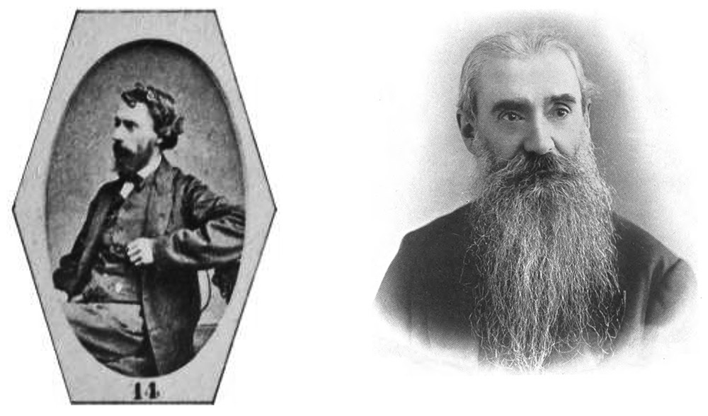
Figure 8. Photographs of Henry Pocklington from ca. 1874 (left) and ca. 1907 (right). The left image is from a collage of 1874 members of the Postal Microscopical Society, which was given to members as a way of “introducing” them to each other. The right image is from the Proceedings of the Insurance Institute of Yorkshire, 1907-1908.
Acknowledgements
Many thanks to Howard Lynk and Peter Paisley for their many
collegial conversations on historical microscopy, especially the countless
discussions that began with “who do you
think made these?”, and to Howard, Peter and Trevor Gillingwater for generous sharing slide images.
Resources
Bracegirdle, Brian (1998) Microscopical Mounts and Mounters, Quekett Microscopical Club, plate 40, slides B and G, pages 184-185
English census, birth, marriage and death records, accessed
through ancestry.co.uk
Hardwicke’s Science-Gossip (1873)
Exchange offer from Henry Pocklington, Vol. 8, page 144
Herapath, William B. (1864) On the genus Synapta, Report of the Thirty-fourth Meeting of the British Association for the Advancement of Science; Held at Bath, September, 1864, pages 97-98
Herapath, William B. (1865) On the genus Synapta, with some new British species, Quarterly Journal of Microscopical Science, new series, Vol. 5, pages 1 - 6
Journal of the Quekett Microscopical Club (1877) Ordinary meeting, Sept. 28, Vol. 5, page 13
Journal of the Royal Microscopical Society (1889) List of members, page lxii
Journal of the Royal Society of Arts (1913) Obituary of Henry Pocklington, Vol. 61
Leading Insurance Men of the British Empire (1892) Henry Pocklington, page 408
Leeds Naturalists’ Club and
Scientific Association, the Sixth Annual Report and President’s Valedictory
Address and a Brief Sketch of the Society’s History, etc. (1876)
pages 8 and 9
List of pen-names so far
determined from English Mechanic and World of Science (accessed
October, 2011) www.englishmechanic.com/pennames.pdf
Monthly Microscopical
Journal: Transactions of the Royal Microscopical Society (1875) H.
Pocklington elected to the RMS, Vol. 13, page 93.
The Naturalist: Journal of the Yorkshire Naturalists’ Union (1878) records of meetings, new series, Vol. 4, pages 64 and 95
The Naturalist: Journal of the Yorkshire Naturalists’ Union (1879) record of Jan. 21 meeting, new series, Vol. 4, page 124
The Naturalist: Journal of the Yorkshire Naturalists’ Union (1882) report of lecture by Henry Pocklington to the Wakefield Naturalist’s and Philosophical Society, new series, Vol. 6, page 152
Obituary Notices of Fellows
of the Royal Society (1953) Henry Cabourn Pocklington, Vol. 8, pages
555-565
The Phrenological Journal
and Science of Health (1888) Report on a lecture by Henry Pocklington on
the microscope as a home help, Vol. 76, pages 157-158
Pocklington, Henry (1867) A stick without end, Hardwicke’s Science-Gossip, Vol. 3, page 187
Pocklington, Henry (1870) The microscope – how to chose it
and how to use it, English Mechanic and
World of Science, Vols. 11, pages 531-532, 578-579, and Vol. 12, pages 4-5,
26-27, 49-50 and 78
Pocklington, Henry (1870) Microscopical jottings in town and
country II, III and IV, English Mechanic
and World of Science, Vol. 11, pages 363, 410 and 436
Pocklington, Henry (1870) Cyclosis in Anacharis, English Mechanic
and World of Science, Vol. 11, pages 553
Pocklington, Henry (1870) numerous brief articles, English Mechanic and World of Science,
Vol. 11, pages 327, 357, 423, 428, 476-477, 478, and 549
Pocklington, Henry (1870) numerous brief articles, English Mechanic and World of Science,
Vol. 12, pages 15-16, 22, 23, 43, 46, 90, 109-110, 135-136, 154, 165, 166, 215,
225-226, 238, 260 and 301
Pocklington, Henry (1870) On the arrangement of a cabinet for
microscopical objects, English Mechanic
and World of Science, Vol. 12, page 289
Pocklington, Henry (1871) numerous brief articles, English Mechanic and World of Science,
Vol. 13, pages 14, 23, 70, 132, 191, 233, 261-262, 285, 331, 367, 382-383, 391
and 463-464
Pocklington, Henry (1871) The optical analysis of bees-wax, The Pharmaceutical Journal, Vol. 2, page 81
Pocklington, Henry (1872) Leaves microscopically considered,
English Mechanic and World of Science,
Vol. 16, pages 279-280 and 352
Pocklington, Henry (1872) The microscope in pharmacy, The Pharmaceutical Journal, Vol. 2,
pages 621, 661, 678-679, 701, 782 and 821
Pocklington, Henry (1872) Some starches microscopically and
polariscopically considered, The Year
Book of Pharmacy, pages 8, 16, 17, 21, 75, 85, 90, etc.
Pocklington, Henry (1872) A chapter in microscopy, Canadian Pharmaceutical Journal, Vol. 5,
page 209
Pocklington, Henry (1872) The microscope in pharmacy, Canadian Pharmaceutical Journal, Vol. 5,
page 309
Pocklington, Henry (1873) Sugar and its adulterations, English Mechanic and World of Science,
Vol. 17, page 189
Pocklington, Henry (1873) numerous contributions on beeswax
and microscopical examination of plants, The
Year Book of Pharmacy, page 555
Pocklington, Henry (1875) The use of optical analysis in
pharmacy, The Year Book of Pharmacy,
page 607
Pocklington, Henry (1871) brief articles, English Mechanic and World of Science, Vol. 22, pages 541 and 565
Post Magazine and Insurance Monitor (1908) Mr. H. Pocklington, Vol. 69, Jan. 11, page 26
Proceedings of the
Insurance Institute of Yorkshire (1908) Picture of Henry
Pocklington inside front cover, and Presidents address on pages 15-26
Report of the British
Association for the Advancement of Science (1883) List of members,
issue 52, page 65
Transactions of the Leeds
Geological Association (1900) List of Members, page 42
Transactions of the Leeds
Geological Association (1914) Report 1913-1914, page 6